Research Article - International Journal of Medical Research & Health Sciences ( 2022) Volume 11, Issue 7
The Potential Risk of Microbial Transmission from Cadavers to its Handlers Including Teaching Staff, Medical Students, Paramedical Staff Working in Dissection Hall
Ashfaq Ul Hassan1, Sajad Hamid1 and Azhar Shfi22Department of Microbiology, SKIMS Medical College, Srinagar, India
Received: 24-May-2022, Manuscript No. ijmrhs-22-65212; Editor assigned: 26-May-2022, Pre QC No. ijmrhs-22-65212(PQ); Reviewed: 09-Jun-2022, QC No. ijmrhs-22-65212; Revised: 23-Jul-2022, Manuscript No. ijmrhs-22-65212(R); Published: 30-Jul-2022, DOI: O
Abstract
Background: Cadavers remain a principal teaching tool for anatomists and medical educators teaching gross anatomy. Infectious pathogens in cadavers that present particular risks include Mycobacterium tuberculosis, hepatitis B and C, the AIDS virus HIV and prions that cause transmissible spongiform encephalopathies such as Creutzfeldt Jakob Disease (CJD) and Gerstmann Straussler Scheinker Syndrome (GSS). It is often claimed that fixatives are effective in inactivation of these agents. Unfortunately cadavers, even though they are fixed, may still pose infection hazards to those who handle them. Specific safety precautions are necessary to avoid accidental disease transmission from cadavers before and during dissection and to decontaminate the local environment afterward. In this brief review, we describe the infectious pathogens that can be detected in cadavers and suggest safety guidelines for the protection of all who handle cadavers against infectious hazards.
Aims: The purpose of this study was to determine if cadavers fixed in a formalin solution are a possible source of microorganisms in the anatomy dissection Hall which can be a potential threat of infection for students.
Material and methods: Students were made to do dissection of routinely preserved cadavers using gloves. Samples from the contaminated gloves were collected and sent to microbiology department for culture and sensitivity.
Results: Using conventional bacteriologic culture and identification methods, our research group was able to successfully recover and identify a variety of organisms from gloves. The results indicate that cadavers processed with 10% buffered formalin have viable organisms on their surfaces that can be a source of contamination. Given the diversity of bacterial species cultured, preserved cadavers used for anatomy education as well as research must be considered a possible source for dissemination of bacterial organisms.
Conclusion: The results indicate that cadavers fixed with formalin are potential source of infection for students as well as faculty. This study underscores protocols to decrease cross contamination.
Keywords
Cadavers, Microorganism, Contamination
Introduction
Human anatomy is a scientific study of structure of human body. The word anatomy is derived from a Greek word “anatome” which means cutting up. Dissection of cadaver is an important pillar and enthusiastic part of MBBS course. Anatomical teaching becomes easy for students with the help of these cadavers and their specimens. First year students of medical profession deal with the cadavers first time. So it is the new experience for them. As cadavers are the main study materials for the students, but can be the source of infections to students and other medical personals who deal with these cadavers during embalming and dissection. The aim of our study was to know and report the various pathogens in fixed cadavers and the proper guidelines which can be put forth before dissection of a cadaver and prevent the transmission of infective agents. Before going for a dissection of a cadaver, one should have a complete knowledge of that cadaver, details of death and about previous hospital records regarding any contagious disease like tuberculosis and HIV, so that more care and precautions could be taken while handling that cadaver.
As we cannot see bacteria with our naked eyes, it is very difficult to know how clean or dirty our hands are. Most of us don’t realize what our hands harbor and how hand washing and hand sanitization can help to prevent so many problems that can occur due to unclean or dirty hands. Every time we touch any object or shake our hands with someone we are most probably picking up bacteria and other viruses too. It has been estimated that we have around 1500 bacteria which are live on every square centimeter of skin of our hands, especially areas under fingernails. Some of the bacteria can be good for us but some can be very dangerous for our health and can cause various infections. Microorganisms have been classified on the basis of relationship they have with humans; commensals organisms that live on our skin, taking benefit from us, but we do not benefit from them. Symbionts the microorganism and humans are mutually beneficial. Pathogens the microorganism benefits but causes disease to the humans. The majority of microorganisms on our skin are normal commensals, as they usually don’t cause any disease. However, in some circumstances commensal microorganisms such as Staphylococcus epidermidis have beneficial as well as pathogenic roles. Sometimes the Commensal microorganisms may prevent growth of pathogenic microorganisms such as Staphylococcus aureus. Most people have similar but not identical microorganisms. Variations can occur due to different age group and different environment.
Dissection is the practical part of anatomy in which students learn from dead body called “cadaver”. Students dissect the body parts and study the structures of body in a detailed manner. It helps them to identify the various structures of body with their own eyes and later they can apply their knowledge to visualize these structures under microscope and also helps them to implement this knowledge for clinical and surgical purposes. But during this handling of the cadaver, students have to be very careful about the contraction of various microbes which can lead to various infections in them. These cadavers are preserved by using fixative agents like Formalin, but researchers have found that this fixative agent is not that effective to inactivate or kill all the microbes on these cadavers. So students are prone to get infected. Not only the students but faculty and other staff are not spared as well [1-3]. The growth of the microbes is inhibited by a procedure called embalming which helps to preserve the cadaver. Fixative agents used in the procedure of embalming are 10% formalin, with added glycerol, water and some disinfectants [4]. It is very important how effective the fixative agent is as the health of the students, faculty and other staff members is at risk. All the people who are directly or indirectly involved with these cadavers are prone to get infected [5].
As our study was mostly concerned with the presence of Staphylococcus, we focused on those bacteria. Staphylococci are gram-positive cocci about 1.0 μm in diameter. They grow in the form of groups or clusters, or pairs or small chains. The shape of these cocci helps us to differentiate between staphylococci from streptococci. The most important test which distinguishes between Staphylococcus from streptococci is the catalase test. Streptococci are catalase negative whereas staphylococci are catalase positive. The test is performed by adding an agar slant or broth culture with multiple drops of 3% hydrogen peroxide. Catalase-positive cultures bubble at once. They are normal habitats present on the skin and mucous membranes, but sometimes can lead to serious infections [6]. Among all the members of staphylococci, Staphylococcus aureus is most pathogenic. Staphylococcus epidermidis is a known cause of various hospital acquired infections (such as prosthetic or indwelling devices), whereas Staphylococci saprophyticus is usually associated with urinary tract infections in young females of reproductive age group. Most common site of entry is skin, but may invade through respiratory tract or urinary tract also. Staphylococcus aureus is responsible for causing boils and abscess, furunculosis, osteomyelitis and endocarditis. Other species of staphylococci (S. lugdunensis, S. haemolyticus, S. warneri, S. schleiferi and S. intermedius) are infrequent pathogens [6]. Staphylococci cohni is a gram positive and coagulase negative member of genus staphylococci which is present in the form of clusters of cocci, it usually present on human skin and is one of the important causes of infections in immunocompromised hosts. The culture of the sample taken from skin shows the presence of diphtherias, staphylococci and various bacilli. Till now it is not proved whether embalming is really effective for killing all the pathogens from the cadavers. The aim of our study was to report presence of any pathogenic bacteria especially staphylococci in cadavers which are used for dissection purposes and how we can prevent transmission of these pathogens to students, faculty and other staff of the dissection hall.
Materials and Methods
Study design: It is a cross-sectional study conducted in February, 2021 in the dissection hall of anatomy department, SKIMS medical college, Bemina.
In our study, first year MBBS students of SKIMS medical college participated after taking consent from all the students. The cadavers which we used in our study were fully formulated as per standard protocol. 120 students were advised to take all the precautions and were asked to use sterile gloves and dissect the body for one hour.
Sample collection: When students did almost the half of dissection, the swabs were collected from the gloves which students were using, by a sterile cotton swab and were kept in Brain Heart Infusion broth (BHI) and were incubated at 37°C overnight.
Processing of samples: All the incubated tubes with swabs were placed in blood agar and MacConkey agar plates and were incubated at 37°C for 24 hours. After that culture plates were studied for any visible growth. The characteristics of the colonies were studied and gram staining was done. Different standard microbiological tests like catalase, clumping factor and tube coagulase test were done to identify different types of cocci. Identification of Staphylococcus aureus was done by tube coagulase, DNase test and mannitol fermentation test. Identification of coagulase negative Staphylococcus species was done as per the methodology explained by IORIO [7].
Statistical analysis: Graph pad prism software, version 3.00 was taken for statistical analysis. The measurements were presented as total number percentage (%), mean and Standard Deviation (SD). P<0.05 was considered as significant.
Result
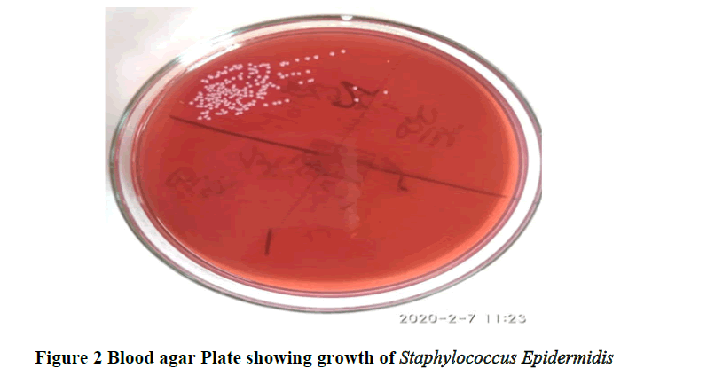
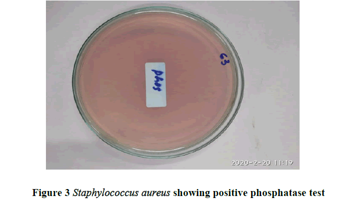
Using conventional bacteriologic culture and identification methods our research group was able to successfully recover and identify a variety of organisms from gloves. The organisms identified in the gloves of students are summarized in the table (Table 1). The results indicate that cadavers processed with 10% buffered formalin have viable organisms on their surfaces that can be a (Figures 1 and 2) source of contamination (Figure 3).
| Micro-organism isolated | Number |
|---|---|
| Staphylococcus aureus | 4 |
| Staphylococcus cohni | 2 |
| Staphylococcus lugdunensis | 3 |
| Staphylococcus saprophyticus | 1 |
| Staphylococcus capitis | 2 |
| Staphylococcus hominis | 2 |
| Staphylococcus warneri | 6 |
| Staphylococcus epidermidis | 3 |
| Nontypeable | 32 |
| Total isolates | 55 |
| Total sample | 120 |
Table demonstrates that 55 types of isolates were obtained in a total of which 32 isolates were non-type able, while as 4 isolates of Staphylococcus aureus were obtained, 2 of Staphylococcus cohni, 3 of Staphylococcus lugdunesis, 1 of Staphylococcus saprophyticus, 2 of Staphylococcus capitis, 2 of Staphylococcus hominis, 6 of Staphylococcus warneri and 3 of Staphylococcus epidermidis.
Discussion
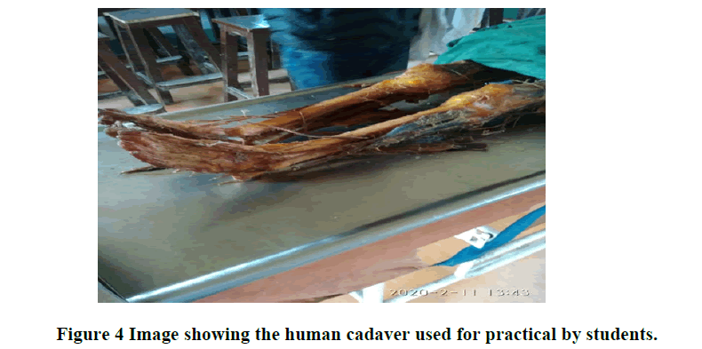
Skin of human beings is the first line of defense against the pathogens and toxins. Skin being the largest organ of the body is continuously affected by internal and external environmental factors. These internal and external factors can change the presence of organisms on skin [8]. Skin microbiology was limited to culture dependent studies, with most samples taken from pathological specimens till recently [9]. However, non-pathological bacteria are detected everywhere on humans, with up to 1 × 10⁷ bacteria per cm2 on the skin [10]. Culture dependent approach being still common, but many microorganisms are very difficult to cultivate and thus remain undetected often. Till date many studies of the human pathogens have focused on the oral and gut microbes. There are various studies on skin microbes but sampling from hands has not been taken into consideration [11,12]. So our knowledge of the microbes present on hands and the factors that influence it is limited and we depend on culture studies only (Figure 4).
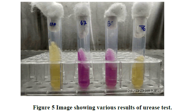
Human hands act as a source of transfer of microorganism from one person to another and from external environment to a person. Hands may harbor many bacteria that are pathogenic, including methicillinresistant Staphylococcus aureus or Escherichia coli; particularly within high risk environments, such as healthcare setting, laboratories and food-handling settings [13]. Use of market products may have an impact on microbes present on hands, with greater pathogen hand carriage on people using a high level of hand hygiene products, while some of the studies have reported decreased level of pathogens while using these products. Frequent hands washing of healthcare workers are colonized with more pathogenic bacteria than those who wash less frequently [14]. Hand washing with soap dispensed from open bulk-refillable dispensers was shown to increase the levels of opportunistic pathogens on children hands in an elementary school [15]. However, many studies have reported the useful results of hand washing and especially use of alcohol-based hand sanitizers for decreasing pathogenic bacteria from hands and thus decreasing the infection rates in different institutional settings. [16,17,18]. The occurrence of pathogens on hands is well-studied; in contrast, hands are rarely considered a source of beneficial bacteria contributing to our healthy micro biome (Figure 5).
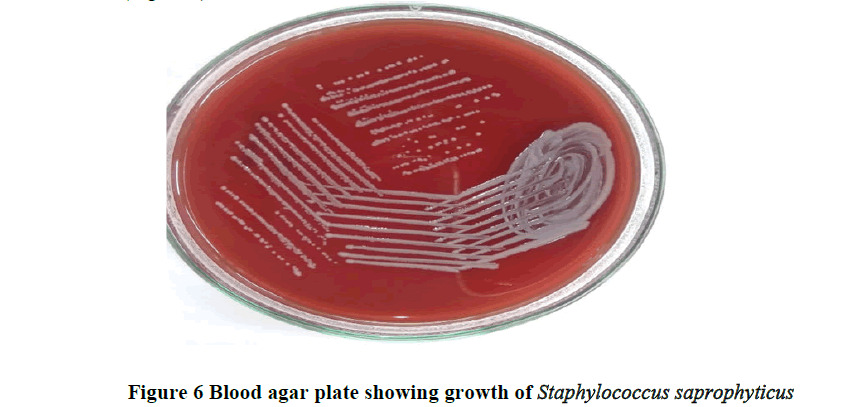
Since hands being essential for intrapersonal and interpersonal exchange of microorganisms, as well as environmental transfer, the changes in the organisms present on hands and various factors influencing them is extremely important [19]. Key topics include understanding the normal microbiome of healthy hands, how microbes are transferred by hands, what factors impact the hand microbiome and whether those impacts are beneficial or detrimental to human health. Like all other occupations, being a member of an anatomy department has its own risks. One of the important and potential sources of infection is the cadaver itself. Cadavers are the main studying materials of anatomists but may have infection risks to people who handle them at the time of embalming and dissection [20]. Infectious pathogens in the cadavers that are present have risks like Mycobacterium tuberculosis, hepatitis B and C viruses, HIV and prions that cause transmissible spongiform encephalopathies [21-27]. The embalming fluid used in anatomy departments contains fixatives, disinfectants, glycerol, salts and water. There is not an adequate data about the efficiency of embalming fluids used commonly. Being the member of anatomy department, one of the important risks of infections is from the cadaver. Special care must be taken to reduce risks to a minimum. Safe working conditions for handling cadavers can be provided through proper education, use of protective clothing and practice of hygienic measures. Dissection laboratory directors must stay up to date on the most recent literature in the field to help ensure the safety of all educators, researchers and students under their charge. In our study we tried to report the presence of any bacteria especially staphylococci in the cadaver used for dissection. A fixative agent used in the process of embalming was 10% formalin with added glycerol, water and some disinfectants (Figure 6).
It was seen in the present study that despite dissecting formalin treated bodies’ variety of micro-organisms were cultured from the samples taken. An inadequate amount of data is available on the antimicrobial abilities of embalming fluids [28]. However from previous studies it has been seen that several disease causing agents may remain viable in cadavers, despite the use of fixative agent [29,30]. But some important pathogens like HIV are killed during embalming of appropriate duration. Few studies have been done to check the disinfecting efficiency of fluids that are used for embalming. Tabaac et al. documented the presence of micro-organisms on formalinembalmed cadavers while Hayashi et al. in their study reported absence of bacteria or yeast like fungi from cadavers embalmed with the same technique [31,32]. The biological risk of fixed cadaver in manipulation and the dissemination of pathogenic microorganisms during anatomy teaching, research and dissection procedures is also reported by some studies. Kabadi et al. also identified Staphylococcus aureus, Enterococcus faecalis and Streptococcus pyogenes by sampling the clothes of students who handled the cadavers [33].
Conclusion
The summary of the present study is that there are viable bacteria recovered from the samples taken from embalmed cadaver. It can be a threat to students, faculty and who use these human cadavers as learning methodology as well as for anatomists and other handlers worldwide who handle these cadavers. It is concluded that present embalming procedures are inadequate for disinfecting of bodies; hence universal precautions should be practiced to prevent infection and cross contamination. Protocols must be developed for safe use of human cadavers in medical learning and Dissection laboratory directors must stay up to date on the most recent literature in the field to help ensure the safety of all educators, researchers and students.
References
- Ramesh G, et al. Detection of microorganisms on formalin fixed and stored pathology tissues: A microbiological study. Journal of oral and maxillofacial pathology: JOMFP, Vol. 21, No. 1, 2017, pp. 64.
- Tabaac B, et al. Bacteria detected on surfaces of formalin fixed anatomy cadavers. Ital J Anat Embryol, Vol. 11, No. 8, 2013, pp. 1-5. 2013, pp. 1-5.
- Morgan DJ, et al. Transfer of Multidrug-Resistant Bacteria to Healthcare Workers’ Gloves and Gowns after Patient Contact Increases with Environmental Contamination. Critical Care Medicine, Vol. 40, No. 4, 2012, pp. 1045-1051.
- O'Sullivan E and Mitchell BS. An improved composition for embalming fluid to preserve cadavers for anatomy teaching in the UK. Journal of anatomy, Vol. 182, No.2, 1993, pp. 295.
- Watson R. General and medical microbiology: summary of biochemical tests. University of Wyoming; 2013.
- Morgan O. Infectious disease risk from dead following natural disasters. Revista panamericana de salud pública, Vol. 15, No. 5, 2004, pp. 307-312.
- Iorio NL et al. Methicillin-resistant Staphylococcus epidermis carrying biofilm formation genes: detection of clinical isolates by multiplex PCR. International Microbiology, Vol. 14, No.1, 2011, pp. 13-17.
- Zeeuwen PL, et al. Microbiome and skin diseases. Current Opinion in Allergy and Clinical Immunology, Vol. 13, No. 5, 2013, pp. 514-520.
- Mathieu A, et al. Life on human surfaces: skin metagenomics. PLos One, Vol. 8, No. 6, 2013, pp. e65288.
- Fredricks DN. Microbial ecology of human skin in health and disease. Journal of Investigative Dermatology Symposium Proceedings, Vol. 6, No. 3, 2001, pp. 167–169.
- Tomic-Canic M, Perez-Perez GI and Blumenberg M. Cutaneous microbiome studies in the times of affordable sequencing. Journal of Dermatological Science, Vol. 75, No. 2, 2014, pp. 82–87.
- Grice E and Segre J, The human microbiome: our second genome. Annual review of genomics and human genetics, Vol. 13, 2012, pp. 151–170.
[Crossref]
- Scott E and Bloomfield SF, The survival and transfer of microbial contamination via cloths, hands and utensils. Journal of Applied Bacteriology, Vol. 68, No. 3, 1990, pp. 271–278.
- Larson E. Skin hygiene and infection prevention: more of the same or different approaches?. Clinical Infectious Diseases, Vol. 29, No. 5, 1999, pp. 1287–1294.
- Zapka CA, et al. Bacterial hand contamination and transfer after use of contaminated bulk-soap refillable dispensers. Applied and Environmental Microbiology, Vol. 77, No. 9, 2011, pp. 2898–2904.
- Fendler EJ, et al. The impact of alcohol hand sanitizer use on infection rates in an extended care facility. American Journal of Infection Control, Vol. 30, No. 4, 2002, pp. 226-233.
- Hilburn J, et al. Use of alcohol hand sanitizer as an infection control strategy in an acute care facility. American Journal of Infection Control, Vol. 31, no. 2, 2003, pp. 109-116.
- White C, et al. The effect of hand hygiene on illness rate among students in university residence halls. American Journal of Infection Control, Vol. 31, No. 6, 2003, pp. 364-370.
- Lax S and Smith DP, Longitudinal analysis of microbial interactions between humans and the indoor environment. Science Magazine, Vol. 345, No. 6200, 2014, pp. 1048-1052.
- Aziz MA, et al. The human cadaver in the age of biomedical informatics. The Anatomical Record: An Official Publication of the American Association of Anatomists, Vol. 269, No. 1, 2002, pp. 20–32.
- Weed LA and Baggenstoss AH. The isolation of pathogens from tissues of embalmed human bodies. American Journal of Clinical Pathology, Vol. 21, No. 12, 1951, pp. 1114-1120.
- Brown P and Gajdusek DC. Survival of scrapie after three years internment. Lancet, Vol. 337, No. 8736, 1991, pp. 269–270.
- Roth D, et al. Detection of hepatitis C virus infection among cadaver organ donors: Evidence for low transmission of disease. Annals of Internal Medicine, Vol. 117, No. 6, 1992, pp. 470–475.
- De Craemer D. Postmortem viability of human immunodeficiency virus-implications for the teaching of anatomy. New England Journal of Medicine, Vol. 331, No. 19, 1994, pp. 1315.
- Healing TD, Hoffman PN and Young SEJ. The infection hazards of human cadavers. Communicable Disease Report. CDR review, Vol. 5, No. 5, 1995, pp. R61–R68.
- Kappel TJ, et al. The viability of Mycobacterium tuberculosis in formalin fixed autopsy tissue: Review of literature and brief report. Hum Pathol, Vol. 27. No. 12, 1996, pp. 1361–1364.
- Cattaneo C, et al. Prevalence of HIV and hepatitis C markers among a cadaver population in Milan. J Clin Pathol, Vol. 52, No. 4, 1999, pp. 267–270.
- Demiryurek, et al. Infective agents in fixed human cadavers: a brief review and suggested guideline. The Anatomical Record: An Official Publication of the American Association of Anatomists, Vol. 269, No. 4, 2002, pp. 194-197.
- De Craemer. Postmortem viability of human immunodeficiency virus implications for the teaching of anatomy. New England Journal of Medicine, Vol. 331, No. 19, 1994, pp. 1315-1316.
- Kaufman M. Dangerous dissections: the hazard from bodies supplied to Edinburgh anatomists, winter session. Journal-Royal College of Physicians of Edinburgh, Vol. 35, No. 3, 2005, pp. 268-274.
- Tabaac B, et al. Bacteria detected on surfaces of formalin fixed anatomy cadavers. Italian Journal of Anatomy and Embryology, Vol. 118, 2013, pp. 1-5.
- Hayashi S, et al. saturated salt solution method: a useful cadaver embalming for surgical skill training. Medicine (Baltimore), Vol. 93, pp. e196.
- Kabadi CJ, et al. Potential pathogen transmission on medical student anatomy laboratory clothing. Medical Student research Journal, 2013, pp. 30-35.


