Research - International Journal of Medical Research & Health Sciences ( 2021) Volume 10, Issue 7
Study of Correlation between hs-CRP and Lipid Profile in Vitamin D Supplemented Hypothyroid Patients
N Ratna Kumari1*, Vijayalakshmi2 and G.S. Prema32Department of Physiology, Saveetha Medical College, Chennai, Tamil Nadu, India
3Department of Physiology, Gandhi Medical College, Hyderabad, Telangana, India
N Ratna Kumari, Department of Physiology, Shadan Institute of Medical Sciences, Hyderabad, Telangana, India, Email: ratnakumari.n.2017@gmail.com
Received: 02-Jun-2021 Accepted Date: Jul 23, 2021 ; Published: 30-Jul-2021, DOI: O
Abstract
Background: Hypothyroidism is a common ailment affecting people globally due to deficiency of thyroid hormones along with their slow metabolism. It is associated with dyslipidemia and inflammation. The hs-CRP (high sensitivity- C-Reactive Protein) is a significant inflammatory marker. A vitamin D deficiency is witnessed in autoimmune diseases and metabolic syndromes. Low vitamin D levels, inflammation, and dyslipidemia were proved to have a connection with autoimmune thyroid diseases. Objectives: Our study aims to estimate the relationship between hs-CRP and lipid profile in vitamin D supplemented hypothyroid patients in comparison to controls and also to compare the abovementioned parameters in without vitamin D supplemented hypothyroid cases. Methods: A cross-sectional study of 6 months period was conducted among 90 subjects attending the General Medicine department of Shadan Institute of Medical Sciences. Based on the inclusion and exclusion criteria, 90 subjects were divided into 3 groups, G-1 (controls=30), G-2 (Vitamin D supplemented hypothyroid subjects=30), and G-3 (without vitamin D supplemented hypothyroid subjects=30). Healthy age and gender-matched euthyroid subjects were taken as controls and patients who were newly diagnosed as hypothyroid, (with increased serum TSH and or with decreased serum T3 or serum T4 levels) were taken as cases. Levels of serum vitamin D, hs-CRP, indicators of thyroid profile (serum TSH, T3, and T4), and indicators of lipid profiles (TC, TAG, HDL, and LDL) were compared between groups. Anthropometric measurements for body mass index were also calculated. Results: 90 subjects participated in our study, the subjects were agematched and female predominance was observed. BMI calculations showed no significant change between the study groups. The hs-CRP levels were improved and found to be statistically significant, in vitamin D supplemented hypothyroid patients. Serum TSH, T3, and T4 levels were within the optimal range. Serum TC mean values were decreased in vitamin D supplemented hypothyroid patients. There was not much difference in HDL levels but an increase in triglycerides and LDL levels with a statistical significance was noted in the vitamin D supplemented hypothyroid patients. When vitamin D supplemented hypothyroid patients and without vitamin D supplemented hypothyroid patients were compared, the TC, TAG, and LDL mean values were decreased and HDL mean value was increased. Between serum TSH and hs-CRP levels of vitamin D supplemented hypothyroid patients, a positive correlation was found, along with statistical significance, when tested by ANOVA. Conclusion: Hypothyroidism is found to be prevalent in females and is associated with mild dyslipidemia. When vitamin D supplemented hypothyroid subjects and without vitamin D supplemented hypothyroid subjects were compared, there was a significant control in hs-CRP levels supporting the advisability of vitamin D supplementation for hypothyroid patients.
Keywords
hs-CRP, Lipid profile, Hypothyroidism, Vitamin D
Abbreviations
T3: Tri-iodothyronine, T4: Thyroxine, TSH: Thyroid Stimulating Hormone, HDL: High-Density Lipoprotein, LDL: Low-Density Lipoprotein, TAG: Triglycerides, TC: Total Cholesterol, hs-CRP: high-sensitivity CReactive ProteinIntroduction
The prevalence of hypothyroidism is around 2% globally [1]. India is in the 2nd place in the world and thyroid disorders are one of the commonest endocrinal ailments in it [2].
Hypothyroidism is a clinical disorder due to deficiency of thyroid hormones, resulting in the reduction of metabolic processes [3]. It affects the various systems of the body, particularly nervous, cardiovascular, pulmonary, reproductive, and renal organ systems [4]. Hypothyroidism is also found to be related to lipid profile; it degrades the lipid synthesis and affects serum lipid levels, specifically LDL and HDL by modifying the gene expression involved in lipid metabolism [5-7].
Vitamin D plays a substantial role in lowering the occurrence of autoimmune diseases and maintenance of vitamin D levels is of much importance in case of deficiency of thyroid hormones [8-10]. Thyroid hormone and vitamin D binds to identical receptors known as steroid hormone receptors. A gene in the Vitamin D receptor was revealed to predispose individuals to autoimmune thyroid diseases [11]. Vitamin D facilitates its effect by binding to VDR, (Vitamin-D Receptor) which leads to the activation of VDR genes, but autoimmune thyroid diseases are associated with VDR gene polymorphism [12,13]. Hence, patients suffering from thyroid problems must try to understand vitamin D’s importance.
Hypothyroid patients suffer from inflammation. hs-CRP (high sensitivity C-Reactive Protein), is an inflammatory marker, vitamin D may have an immunomodulatory effect in hypothyroid patients, which could be related to systemic hs-CRP [14].
In our study, we tried to find out a correlation between hs-CRP and lipid profile in vitamin D supplemented hypothyroid patients.
Materials and Methods
A cross-sectional study of 6 months period was conducted among 90 subjects attending the General Medicine department of Shadan Institute of Medical Sciences. The study comprised of subjects of either gender, aged between 15-45 years. All the subjects were instructed to fill informed consent form before enrolment in the study. An ethical committee permission letter was taken from the institution before the commencement of the study.
Group Allocation
90 subjects were divided into 3 groups
Group-1=Healthy controls (n=30)
Group-2=Vitamin D supplemented hypothyroid subjects (n=30)
Group-3=Without Vitamin D supplemented hypothyroid subjects (n=30)
Inclusion Criteria
Controls: Healthy age and gender-matched euthyroid subjects.
Cases: Newly diagnosed hypothyroid subjects (with increased serum TSH and or with decreased T3 or serum T4 levels).
Exclusion Criteria
Subjects are already on hypothyroid medication. Subjects suffering from any cardiovascular pulmonary, renal, neurological, or reproductive disorders.
Investigations Done
Thyroid profile: Done by Chemiluminescence Immunoassay (CLIA) [15].
hs-CRP: Done by Immunoturbidimetric assay [16].
Lipid profile: Done by enzymatic colourimetric methods and calculations [17].
Vitamin D: Done by Mini vidas automated immunoassay analyzer [18].
Anthropometric measurements for body mass index were also calculated.
Five ml of blood sample was collected from all the subjects for estimation of biochemical parameters. Serum was separated from collected blood and free T3, free T4, and TSH were assayed by using the Chemiluminescence Immunoassay (CLIA) method. The lipid profile was estimated by the enzymatic colorimetric method. Quantitative assay of vitamin D levels and high-sensitivity C-Reactive Protein (hs-CRP), was done by using Mini vidas automated immunoassay analyzer and immunoturbidimetric assay, respectively.
Statistical Analysis
Executed by using the SPSS software. Data analysis was done by statistical tools, a) Descriptive analysis, b) student t-test, c) Chi-square test, d) Pearson’s correlation and e) ANOVA.
Results
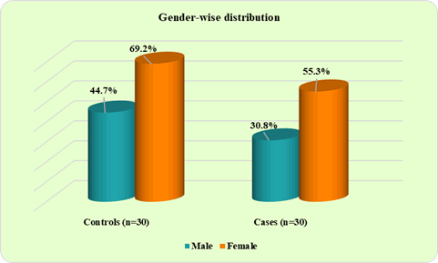
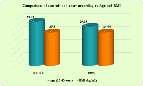
90 subjects who participated in our study, were age-matched (mean age 33 years) and female predominance was observed (controls=69.2% and cases=55.3%) (Figure 1). Figure 2 depicts the comparison of G-1 and G-2 according to age and BMI. The mean age of controls (G-1) and cases (G-2) was found to be 33.87 ± 5.65 and 29.93 ± 8.86 years, respectively and BMI values didn’t show much difference between controls and cases subjects of G-1 (25.50 ± 2.39) and G-2 (24.04 ± 4.76), respectively as well as in G-2 (24.04 ± 4.76) and G-3 (24.73 ± 4.95) patients there was no significant change (Table 1).
| Parameters | G-2 | G-3 | p-value |
|---|---|---|---|
| (Vitamin D supplemented hypothyroid subjects) (n=30) | (Without Vitamin D supplemented hypothyroid subjects) (n=30) | ||
| BMI (kg/m2) | 24.04 ± 4.76 | 24.73 ± 4.95 | 0.072 |
| Serum Vitamin D (ng/ml) levels | 27.80 ± 4.20 | 17.60 ± 5.12 | 0.176 |
| Serum TSH (mµ/L) levels | 4.81 ± 4.60 | 5.45 ± 2.33 | 0.276 |
| Serum T3 (pg/ml) levels | 2.11 ± 0.88 | 2.29 ± 1.51 | 0.001* |
| Serum T4 (ng/dl) | 6.49 ± 2.04 | 6.77 ± 1.96 | 0.001* |
| TC levels | 188.83 ± 39.03 | 238.97 ± 37.99 | 0.069 |
| TAG levels | 139.80 ± 30.22 | 169.57 ± 50.48 | 0.839 |
| HDL levels | 37.40 ± 6.15 | 30.80 ± 4.62 | 0.126 |
| LDL levels | 87.83 ± 19.82 | 100.37 ± 31.63 | 0.001* |
| hs-CRP levels | 3.43 ± 1.36 | 7.27 ± 1.96 | 0.001* |
Data represented as Mean ± SD, *: represents statistical significance, p-value <0.001 is significant. BMI: Body Mass Index, TSH: Thyroid Stimulating Hormone, T3; Tri-iodothyronine and T4: Thyroxine, TC: Total Cholesterol, TAG: Triglycerides, HDL: High-Density Lipoprotein, LDL: Low-Density Lipoprotein, hs-CRP: high sensitivity C-Reactive Protein
Table 2 represents comparison of thyroid profile, lipid profile, and hs-CRP levels between controls (G-1) and cases (G- 2) subjects. The mean serum TSH (4.81 ± 4.60), T3 (1.53 ± 0.28), and T4 (6.49 ± 2.04) levels were within the optimal range in the cases (G-2) when compared with controls (G-1). Serum TC mean values were decreased in the cases subjects (G-2). There was not much difference in HDL levels but an increase in TAG (Triglycerides) (139.80 ± 30.22) and LDL levels (87.83 ± 19.82) with a statistical significance (p<0.001) was noted in the cases subjects (group-2). The hs-CRP levels were considerably improved (3.43 ± 1.36) and found to be statistically significant (p<0.001) in cases subjects (G-2) (Table 2). Distribution of controls (group-1) and cases subjects (G-2) according to their hs-CRP levels are shown in Table 3 where, hs-CRP levels were in improved percentage (84.6%) and is statistically significant (p-value<0.001) with Chi-square value-21.991*, in cases subjects (G-2).
| Parameters | G-1 | G-2 | p-value |
|---|---|---|---|
| Controls (n=30) | Cases (with vitamin D supplementation) (n=30) | ||
| Serum TSH (mµ/L) levels | 4.78 ± 4.67 | 4.81 ± 4.60 | 0.001* |
| Serum T3 (pg/ml) levels | 1.18 ± 0.27 | 1.53 ± 0.28 | 0.152 |
| Serum T4 (ng/dl) | 6.04 ± 1.64 | 6.49 ± 2.04 | 0.127 |
| TC levels | 196.93 ± 24.61 | 188.83 ± 39.03 | 0.116 |
| TAG levels | 109.40 ± 16.88 | 139.80 ± 30.22 | 0.001* |
| HDL levels | 37.10 ± 6.33 | 37.40 ± 6.15 | 0.167 |
| LDL levels | 74.23 ± 12.30 | 87.83 ± 19.82 | 0.001* |
| hs-CRP levels | 1.70 ± 0.70 | 3.43 ± 1.36 | 0.001* |
Data represented as Mean ± SD, SD: Standard Deviation, * represents statistical significance, p-value <0.001 is significant
| hs-CRP | G-1 | G-2 |
|---|---|---|
| Controls (n=30) | Cases (with vitamin D supplementation) (n=30) | |
| <3mg/l | 26 (76.5%) | 8 (23.5%) |
| ≥ 3mg/l | 4 (15.4%) | 22 (84.6%) |
| Total | 30 (100%) | 30 (100%) |
hs-CRP (high sensitivity C-Reactive Protein) Chi square value=21.991*, p-value=0.001*
In Table 4, between serum TSH and hs-CRP levels of G-2 case subjects, a positive correlation was found (r-0.269*, p<0.001), along with statistical significance (p<0.001), when tested by ANOVA (Table 5). When group-2 (Vitamin D supplemented hypothyroid subjects) and group 3 (without Vitamin D supplemented hypothyroid subjects) were compared, the TC, TAG, and LDL (p<0.001) mean values were decreased and HDL mean value was increased in cases subjects (G-2, Vitamin D supplemented hypothyroid subjects). Serum vitamin D levels have significantly improved in cases subjects (G-2). No prominent difference was found in the thyroid profile of G-2 and G-3 subjects.
| Parameters | r-value | p-value |
|---|---|---|
| TSH vs. hs-CRP (Group-2, Cases with vitamin D supplement) | 0.269* | 0.001* |
| TSH: Thyroid Stimulating Hormone; hs-CRP (high sensitivity C-Reactive Protein) | ||
| Parameters | G-1 | G-2 | G-3 | Total | p-value |
|---|---|---|---|---|---|
| Controls (n=30) | (Vitamin D supplemented hypothyroid subjects) (n=30) | (Without Vitamin D supplemented hypothyroid subjects) (n=30) | |||
| Serum TSH (mµ/L) levels | 2.90 ± 0.83 | 4.81 ± 4.60 | 5.45 ± 2.33 | 4.38 ± 3.17 | 0.0004* |
| hs-CRP levels | 1.70 ± 0.70 | 3.43 ± 1.36 | 7.27 ± 1.96 | 4.13 ± 2.73 | 0.0001* |
colspan="6">Data represented as Mean ± SD; *: represents statistical significance, p-value<0.001 is significant. TSH: Thyroid Stimulating Hormone; hs-CRP: high sensitivity C-reactive protein
Discussion
The whole purpose of our entire study was to analyze the correlation between inflammatory marker, hs-CRP (high sensitivity C-reactive protein) and lipid profile in vitamin D supplemented hypothyroid patients.
In our study, a total of 90 subjects participated. Out of 90 subjects, healthy age and gender-matched euthyroid subjects were taken as controls, and patients who were newly diagnosed as hypothyroid, (with increased serum TSH and or with decreased serum T3 or T4 levels) were taken as cases.
Both the controls and cases were age-matched (mean age 33 years) and female predominance was observed (controls=69.2% and cases=55.3%). In a similar study reported by Morganti S, et al., a higher prevalence rate of hypothyroidism in women with advancing age was detected [19]. The study by Unnikrishnan A, et al., on hypothyroid patients, has revealed that the female gender has a significant association with hypothyroidism [20].
The relationship between obesity and hypothyroidism has been studied for decades, in our study comparison of controls and cases subjects BMI was done. It was revealed, that the BMI values of controls (G-1) (25.50 ± 2.39) and cases (G-2) (24.04 ± 4.76) subjects were not of much difference. When BMI calculations between vitamin D supplemented hypothyroid subjects (24.04 ± 4.76) and without vitamin D supplemented hypothyroid subjects (24.73 ± 4.95) were compared, there was not much change in both the groups. Few studies inconclusively established the above result, work done by Ríos-Prego, Monica, et al., and Talaei A, et al., witnessed that BMI is not strongly influenced by vitamin D in thyroid dysfunction [21,22].
hs-CRP (high sensitivity C-reactive protein), being an inflammatory marker, is closely related to hypothyroidism. In our study, the hs-CRP levels were improved (3.43 ± 1.36) and found to be statistically significant (p<0.001), in vitamin D-supplemented hypothyroid subjects. Between TSH and hs-CRP levels of vitamin D supplemented hypothyroid subjects, a positive correlation was found (r-0.269*, p<0.001), along with statistical significance (p<0.001), when tested by ANOVA was found. Vitamin D’s role in immune cell functions and inflammation, cannot be denied with these results. It is in agreement with a study done by Mirhosseini N, et al., where, improved vitamin D levels in hypothyroid patients affected hs-CRP levels and reduced inflammation indicating the importance of maintaining vitamin D levels in hypothyroid patients [23].
In our study, a prominent difference was not noted in serum TSH, T3, and T4 levels of vitamin D supplemented hypothyroid subjects compared to those without vitamin D supplemented hypothyroid subjects. Serum TSH, T3, and T4 levels were within the optimal range, in controls and cases study groups. A similar result was observed by Simsek Y, et al., where thyroid function tests did not show significant change with Vitamin D therapy in study groups [24].
Thyroid dysfunctions are known to affect lipid metabolism, dyslipidemia has an eminent relation with thyroid dysfunction [25]. In our study, total cholesterol levels were decreased in vitamin D supplemented hypothyroid subjects which is in agreement with the study done by Husham I, et al., where, the study demonstrated that vitamin D improved serum levels of total cholesterol [26]. There was an increase in the levels of triglycerides and LDL but no change in HDL levels in vitamin D supplemented hypothyroid subjects (G-2) when compared with controls. Similar findings were reported in the study done by Kshetrimayum V, et al., in which a significant increase in TAG and LDL was determined [27]. When vitamin D supplemented hypothyroid subjects (G-2) and without vitamin D supplemented hypothyroid subjects (G-3) were compared, the TC, TAG, and LDL (p<0.001) mean values were decreased and HDL mean value was increased. Mansorian B, et al., also reported the same results in their study, improved HDL levels and decreased TC, TAG, and LDL detection through their study was proved [28].
Conclusion
The results in our study show that hypothyroidism is found to be prevalent in females and is associated with mild dyslipidemia. When vitamin D supplemented hypothyroid subjects and without vitamin D supplemented hypothyroid subjects were compared, there was a significant control in hs-CRP levels supporting the advisability of vitamin D supplementation for hypothyroid patients. Maintenance of vitamin D levels is recommended for all patients suffering from hypothyroidism.
References
- Vanderpump, Mark PJ. "The epidemiology of thyroid disease." British Medical Bulletin, Vol. 99, No. 1, 2011, pp. 39-51.
- Kochupillai, N. "Clinical endocrinology in India." Current Science, Vol. 79, No. 8, 2000, pp. 1061-67.
- Gardner, David G., and Francis Sorrel Greenspan, eds. "Basic & clinical endocrinology." McGraw-Hill, 2007.
- Shahid, Muhammad A., Muhammad A. Ashraf, and Sandeep Sharma. "Physiology, thyroid hormone." StatPearls Publishing, 2018.
- Hariharan, Somasundaram, et al. "Dyslipidemia in hypothyroid subjects with Hashimoto’s thyroiditis." International Journal of Medical Science and Public Health, Vol. 4, No. 9, 2015, p. 1.
- Liu, Yan-Yun, and Gregory A. Brent. "Thyroid hormone crosstalk with nuclear receptor signaling in metabolic regulation." Trends in Endocrinology & Metabolism, Vol. 21, No. 3, 2010, pp. 166-73.
- Rizos, C. V., M. S. Elisaf, and E. N. Liberopoulos. "Effects of thyroid dysfunction on lipid profile." The Open Cardiovascular Medicine Journal, Vol. 5, 2011, pp. 76-84.
- Baeke, Femke, et al. "Vitamin D: Modulator of the immune system." Current Opinion in Pharmacology, Vol. 10, No. 4, 2010, pp. 482-96.
- Subashree, R., and Radhika Arjunkumar. "Vitamin D deficiency in periodontal health." Research Journal of Pharmacy and Technology, Vol. 7, No. 2, 2014, pp. 248-52.
- Mackawy, Amal Mohammed Husein, Bushra Mohammed Al-Ayed, and Bashayer Mater Al-Rashidi. "Vitamin D deficiency and its association with thyroid disease." International Journal of Health Sciences, Vol. 7, No. 3, 2013, pp. 267-75.
- Friedman, Theodore C. "Vitamin D deficiency and thyroid disease." Good Hormone Health, 2011.
- Pike, J. Wesley, and Mark B. Meyer. "The vitamin D receptor: New paradigms for the regulation of gene expression by 1, 25-dihydroxyvitamin D3." Rheumatic Disease Clinics, Vol. 38, No. 1, 2012, pp. 13-27.
- Ban, Yoshiyuki, Matsuo Taniyama, and Yoshio Ban. "Vitamin D receptor gene polymorphism is associated with Graves’ disease in the Japanese population." The Journal of Clinical Endocrinology & Metabolism, Vol. 85, No. 12, 2000, pp. 4639-43.
- Pearce, Elizabeth N., et al. "The prevalence of elevated serum C-reactive protein levels in inflammatory and noninflammatory thyroid disease." Thyroid, Vol. 13, No. 7, 2003, pp. 643-48.
- Bhatt, Mahendra Prasad, et al. "A multi-center assessment of thyroid function test precision in Chemiluminescence Immunoassay (CLIA) systems." Journal of Gandaki Medical College-Nepal, Vol. 11, No. 2, 2018, pp. 8-13.
- Dupuy, Anne M., et al. "Immunoturbidimetric determination of C-reactive protein (CRP) and high-sensitivity CRP on heparin plasma. Comparison with serum determination." Clinical Chemistry and Laboratory Medicine, Vol. 41, No. 7, 2003, pp. 948-49.
- Mizoguchi, Toshimi, Toshiyuki Edano, and Tomoyuki Koshi. "A method of direct measurement for the enzymatic determination of cholesteryl esters." Journal of Lipid Research, Vol. 45, No. 2, 2004, pp. 396-401.
- Moreau, Emmanuel, et al. "Performance characteristics of the VIDAS® 25-OH Vitamin D Total assay-comparison with four immunoassays and two liquid chromatography-tandem mass spectrometry methods in a multicentric study." Clinical Chemistry and Laboratory Medicine (CCLM), Vol. 54, No. 1, 2016, pp. 45-53.
- Morganti, S., et al. "Thyroid disease in the elderly: Sex-related differences in clinical expression." Journal of Endocrinological Investigation, Vol. 28, No. 11 Suppl Proceedings, 2005, pp. 101-04.
- Unnikrishnan, Ambika Gopalakrishnan, et al. "Prevalence of hypothyroidism in adults: An epidemiological study in eight cities of India." Indian Journal of Endocrinology and Metabolism, Vol. 17, No. 4, 2013, pp. 647-52.
- Ríos-Prego, Monica, Luis Anibarro, and Paula Sanchez-Sobrino. "Relationship between thyroid dysfunction and body weight: A not so evident paradigm." International Journal of General Medicine, Vol. 12, 2019, pp. 299-304.
- Talaei, Afsaneh, Fariba Ghorbani, and Zatollah Asemi. "The effects of Vitamin D supplementation on thyroid function in hypothyroid patients: A randomized, double-blind, placebo-controlled trial." Indian Journal of Endocrinology and Metabolism, Vol. 22, No. 5, 2018, pp. 584-88.
- Mirhosseini, Naghmeh, et al. "Physiological serum 25-hydroxyvitamin D concentrations are associated with improved thyroid function-observations from a community-based program." Endocrine, Vol. 58, No. 3, 2017, pp. 563-73.
- Simsek, Yasin, et al. "Effects of Vitamin D treatment on thyroid autoimmunity." Journal of Research in Medical Sciences: The Official Journal of Isfahan University of Medical Sciences, Vol. 21, 2016.
- Mohammed, Sayed, et al. "Evaluation of serum lipid profile in Sudanese patient with thyroid dysfunction" Scholars Journal of Applied Medical Sciences (SJAMS), Vol. 3, No. 6A, 2015, pp. 2178-82.
- Al-mafraji, Emad Hisham Ahmed, and Rafah Razooq Hameed Al-Samarrai. "Evaluation the correlation between Vitamin D deficiency and lipid profile in thyroid disease." Journal of Al-Nisour University College, Vol. 5, 2018, pp. 246-63.
- Kshetrimayum V, Usha S M R, Vijayalakshmi P. "A study of hs-CRP and lipid profile in hypothyroid adults at tertiary care hospital." International Journal of Clinical Biochemistry and Research, Vol. 6, No. 3, 2019, pp. 303-10.
- Mansorian, Behnam, et al. "Serum vitamin D level and its relation to thyroid hormone, blood sugar and lipid profiles in Iranian sedentary work staff." Hospital Nutrition: Official organ of the Spanish Society of Parenteral and Enteral Nutrition, Vol. 35, No. 5, 2018, pp. 1107-14.


