Case Reports - International Journal of Medical Research & Health Sciences ( 2023) Volume 12, Issue 3
Smile Designing: A Multidisciplinary Approach for Dental Aesthetics
Tamilselvan Kumar*Tamilselvan Kumar, Department of Periodontics, Rajas Dental College and Hospital, Tirunelveli, Tamil Nadu, India, Email: tamilbdstirupur@gmail.com
Received: 17-Oct-2022, Manuscript No. IJMRHS-22-77511; Editor assigned: 19-Oct-2022, Pre QC No. IJMRHS-22-77511 (PQ); Reviewed: 02-Nov-2022, QC No. IJMRHS-22-77511; Revised: 09-Mar-2023, Manuscript No. IJMRHS-22-77511 (R); Published: 16-Mar-2023
Abstract
Smile is the ability of a person to express his/her emotion with the structure and movement of the teeth and lips. Smile determines how well a person can function in the society. Correct dental proportion is essential in creating an esthetically pleasing smile. Central incisors are considered the key and the most dominant teeth in the smile and they should display pleasing proportions. In a perfect smile, the gum line follows the upper lip or is just above it and ensures that just enough gums (2 mm-3 mm) are exposed. This case report depicts the successful management of a patient who reported to the department of periodontics with the chief complaint of shortened clinical crown length and spacing between teeth in the upper anterior teeth region. On clinical examination, there was shortened clinical crown height with excessive gingival display in upper anterior and midline diastema was noted. Gingivectomy was performed to increase the clinical crown length and to reduce the overexposed gingival tissue. Laminate veneers were used to correct the midline diastema.
Keywords
Smile designing, Midline diastema, Crown lengthening, Gingivectomy, Laminate veneer
Introduction
In this modern era of dentistry, patients demand for highly esthetic treatment outcome has increased significantly. Innovations in the field of dentistry have shifted the way people think about their dental aesthetics [1]. Individual personality is highly influenced by their facial and dental appearances [2]. Architecting a beautiful smile requires the interdisciplinary approach in the field of dentistry. Dentists should be capable of skillfully blending science and art to sculpt a more attractive smile. The presence or absence of teeth, its alignment in the arch, gingival exposure, diastema and the amount of display both horizontally and vertically are the factors which lead to the esthetic perception of a beautiful smile.
The gingival exposure between the gingival margin of anterior central incisors and the inferior border of upper lip while smiling is termed as normal gingival display. Excessive gingival display of more than 2 mm is termed as gummy smile. Gummy smiles range from mild, moderate and advanced to severe. Mazzuco classified gummy smile into anterior, posterior, mixed or asymmetric based on the excessive contraction of muscles involved [3]. Surgical correction of the overgrown tissues is frequently necessary to accomplish an esthetic and functional outcome. The treatment consists of scalpel gingivectomy or laser gingivectomy.
Laser is one of the most promising new technical modalities in periodontal treatment [4]. However, some laser wavelengths work on both hard and soft tissues (2,780 nm and 2,940 nm) while other lasers, such as the 810 nm diode work on only soft tissues and have a very good surgical and hemostatic action on soft tissues following maxillary vestibular frenectomies, gingivectomies, recontouring of gingival overgrowth, surgical exposure of buccally/palatally placed teeth and operculectomies. Also, the soft tissue diode lasers have an excellent incision performance with a cutting depth of 2 mm-6 mm and have an added advantage over conventional surgery in that there is a sealing of small blood and lymphatic vessels resulting in haemostasis and reduced postoperative edema. Target tissues are also disinfected as a result of local heating and production of an eschar layer and a decreased amount of scarring due to decreased postoperative tissue shrinkage.
Space between the teeth is termed as diastema and it affects the aesthetics of an individual. Even though orthodontic treatment can correct certain problems, patients having minor esthetic problems are not willing for orthodontic treatment because of its time consuming process. The role of periodontist and other dental specialist in recreating smile in these situations becomes critical. One of the preferred treatment options for these problems includes preparation of thin shells of ceramics known as Porcelain Laminate Veneers (PLV) which can be bonded to the facial surface of anterior teeth using recent bonding agents and dual cure cements [5]. This case report depicts the successful management of an unaesthetic smile due to the presence of gummy smile and diastema by the interdisciplinary approach [6].
Case Presentation
Anamnesis and clinical examination
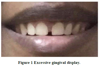
A 20 years old female patient came to the department of periodontology and implantology, Komarapalayam, reporting discontentment with the visual aspect of her smile which exhibited great exposure of the gingiva during her smile with short teeth and midline diastema as shown in Figures 1 and 2. In the anamnesis, it was noted that the patient had no systemic abnormalities. No abnormality was detected in the extra oral examination [7]. In the intra oral examination, presence of gummy smile associated with the presence of keratinised mucosa covering the anatomical crown was observed.
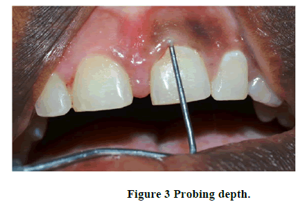
Midline diastema was seen between two upper central incisors. There was no bleeding on probing and the probing depth was ranging from 2 mm to 3 mm as shown in Figure 3. The width of keratinised mucosa was ranging from 5 mm to 10 mm throughout the upper anterior sextant. Treatment plan in the upper arch from right lateral incisor to the left lateral incisor was performed based on the amount of gingiva to be removed in order to offer more precision and safety to the surgical procedure [8].
Surgical procedure
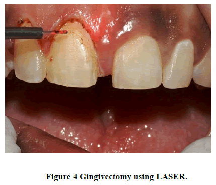
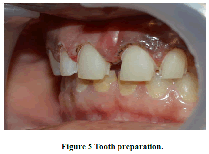
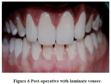
The surgical procedure started with extra oral antisepsis with 2% chlorhexidine in the lower and middle thirds of the face and intraoral antisepsis with 0.12% chlorhexidine. Local anaesthesia last performed by giving bilateral infraorbital nerve block with 2% lignocaine with 1:80,000 adrenaline. Biological width was recorded using transgingival probing. Gingival marking is done after placing stent over the teeth. Gingivectomy was done using laser (Figure 4). Patient was asked to report after 1 week for review and tooth preparation. On review the gingival shape, contour, scalloping and margin were regained. Tooth preparation was done in relation to 11, 12, 21 and 22 for laminate veneers as shown in Figure 5. Prepared tooth were etched using 37% phosphoric acid, bonding agent was applied followed by which laminate veneers were fixed using composite resin (Figure 6).
Discussion
The extent of gingival exposure when smiling is an important factor which can affect the aesthetics. Generally, an ideal smile is one that exposes the minimum gingiva [9]. The literature shows that a smile can be classified as gingival when the cervico incisal height of the teeth is completely seen or when the amount of visible gingival tissue reaches values greater than 3 mm. Excessive gingival display is a clinical finding with many etiologies and may include extra or intraoral components. It is important to identify the type of gummy smile to establish correct treatment. Gingivectomy can be performed by conventional scalpels, electro surgery, chemosurgery and laser. For many intra oral soft tissue surgical procedures, the laser is a viable alternative to the conventional techniques. The commercially available dental instruments have emission wavelengths ranging from 488 nm-10,600 nm and are all non-ionizing radiations which don’t cause any mutations. Advantages of lasers include increased coagulation that yields a dry surgical field and better visualization; the ability to negotiate curvatures and folds within tissue contours; tissue surface sterilization, reduction in bacteraemia, decreased edema, swelling and scarring, decreased pain, faster healing response and increased patience acceptance [10]. Diode laser has a broad range of applications, which includes tissue retraction during restorative procedures, gingivectomy, gingivoplasty, crown lengthening, frenectomy, bacterial decontamination and removal of diseased epithelial lining during periodontal treatments [11]. Porcelain Laminate Veneers (PLVs) have become the alternative to composite restorations ceramic crowns and the traditional porcelain fused to metal [12]. The estimated survival probability of porcelain laminate veneers over a period of 10 years is 91%. Aesthetics is adversely affected by diastemas and the technique followed in this case report may be used to correct a wide range of mid line diastema without compromising the aesthetics and stability of treated outcome.
Conclusion
The treatment of gummy smile requires proper treatment planning. This case report depicts the treatment of excessive gingival display effectively by gingivectomy using diode laser with minimal to no discomfort postoperatively resulting in better gingival shape, contour, scalloping and gingival margin. The recovery of function and smile aesthetics of the patient with porcelain laminate veneers allowed excellent results along with conservative tooth preparations.
Conflicts of Interest
There are no conflicts of interest.
References
- Mushannavar LS. Smile designing-A case report. The Journal of the Indian Prosthodontic Society, Vol. 18, No. 2, 2018, pp. 98.
[Crossref] [Google Scholar] [PubMed]
- Raghu R, et al. Smile rejuvenation: A case report. Journal of Conservative Dentistry, Vol. 17, No. 5, 2014, pp. 495-498.
[Crossref] [Google Scholar] [PubMed]
- Mazzuco R and Hexsel D. Gummy smile and botulinum toxin: A new approach based on the gingival exposure area. Journal of The American Academy of Dermatology, Vol. 63, No. 6, 2010, pp. 1042-1051.
[Crossref] [Google Scholar] [PubMed]
- Ize-iyamu INS, Saheeb BD and Edetanlen BE. Comparing the 810 nm diode laser with conventional surgery in orthodontic soft tissue procedures. Ghana Medical Journal, Vol. 47, No. 3, 2013, pp. 107-111.
[Google Scholar] [PubMed]
- Miara P. Esthetic dentistry and ceramic restorations. 1st edition, Taylor and Francis group, New York, 2013;161-214.
- Dumfahrt H and Schäffer H. Porcelain laminate veneers. A retrospective evaluation after 1 to 10 years of service: Part II-clinical results. The International Journal of Prosthodontics, Vol. 13, No. 1. 2000, pp. 9-18.
[Google Scholar] [PubMed]
- Frese C, Staehle HJ and Wolff D. The assessment of dentofacial esthetics in restorative dentistry: A review of the literature. The Journal of the American Dental Association, Vol. 143, No. 5, 2012, pp. 461-466.
[Crossref] [Google Scholar] [PubMed]
- Antoniazzi RP, et al. Impact of excessive gingival display on oral health related quality of life in a Southern Brazilian young population. Journal of Clinical Periodontology, Vol. 44, No. 10, 2017, pp. 996-1002.
[Crossref] [Google Scholar] [PubMed]
- Goldman L. Chromophores in tissue for laser medicine and laser surgery. Lasers in Medical Science, Vol. 5, No. 3, 1990, pp. 289-292.
- Oiveira PS, et al. Aesthetic surgical crown lengthening procedure. Case Reports in Dentistry, 2015; Vol. 2015, 2015, pp. 10-13.
[Crossref] [Google Scholar] [PubMed]
- Viswambaran M, Londhe SM and Kumar V. Conservative and esthetic management of diastema closure using porcelain laminate veneers. Medical Journal Armed Forces India, Vol. 71, No. 2, 2015, pp. 581-585.
[Crossref] [Google Scholar] [PubMed]
- Frese C, Staehle HJ and Wolff D. The assessment of dentofacial esthetics in restorative dentistry: A review of the literature. The Journal of the American Dental Association, Vol. 143, No. 5, 2012, pp. 461-466.
[Crossref] [Google Scholar] [PubMed]

