Case Report - International Journal of Medical Research & Health Sciences ( 2023) Volume 12, Issue 5
Rare Demographic Presentation of Epidermoid Cysts in Postauricular Region: A Case Series
Smit Shah*, Vaishali Sangole, Jubina PP, Janvi Kansagra and Neha DeshmukhSmit Shah, Department of Dermatology, University of Delhi, New Delhi, India, Email: smitshah436@gmail.com
Received: 21-Dec-2022, Manuscript No. IJMRHS-22-84389; Editor assigned: 23-Dec-2022, Pre QC No. IJMRHS-22-84389 (PQ); Reviewed: 06-Jan-2023, QC No. IJMRHS-22-84389; Revised: 17-Mar-2023, Manuscript No. IJMRHS-22-84389 (R); Published: 24-Mar-2023
Abstract
Epidermoid cysts are benign lesions encountered throughout the body. Eighty percent of epidermoid cyst seen in ovaries and testicles, whereas in head and neck region they account for only 1.6%-7.0%. 1.6% of epidermoid cysts occur in oral cavity and they account for 0.01% of all the oral cavity cysts. Although epidermoid cysts are rarely encountered in the oral cavity, the possibility that they may occur warrants the need for successful management to avoid misdiagnosis.
Keywords
Epidermoid cyst, Encountered, Misdiagnosis, Ovaries and testicles, Oral cavity
Introduction
Compared with other cutaneous cysts of the head and neck region, postauricular epidermoid cysts are rare, asymptomatic benign swellings. Epidermoid cysts are developmental cutaneous cysts derived from trapped pouches of ectodermal, neural tube and surface ectodermal separation failure and from traumatic implantation of squamous epithelium. Only 7% epidermoid cyst occurs in the head and neck region. For site distribution of these cysts, postauricular location is a very rare occurrence. Most common out of the two is the dermoid type. Because of the location and the painless nature of the cyst, patients may take a long time to seek medical intervention [1].
Aims and objectives: The aim is to describe the clinical profile of postauricular epidermoid cysts. The objectives are to describe the demographic characteristic, symptoms duration and histological diagnosis in patients.
Case Presentation
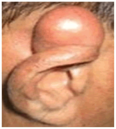
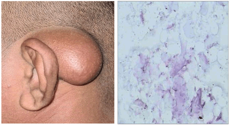
A descriptive study was conducted in 5 patients between 1-18 yrs. in a period of 1 year. The clinical records were analyzed for age, sex, duration, histological types, postauricular side involved (right or left), symptom(s) presentation, reasons for seeking medical intervention and the recurrence after treatment (Figure 1) [2]. Inclusion criteria included patients between 1-18 years with postauricular epidermoid cyst. Excluded from this study were patients older than 18 years and the other developmental cyst(s) in the other sites of the head and neck region. Data were analyzed by simple statistical analysis. On clinical examination of all the patients, swelling was soft, cystic, globular and non-tender with restricted motility. Margins were well defined. Skin over the swelling was normal and not attached to it. There was no discharging sinus or pointing abscess. Bruit or any pulsations were absent. There was no history trauma, fever, loss of appetite, discharging ear, difficulty in hearing, etc. The patient had no other complaints or other constitutional symptoms [3]. There was no history of such lesions in his family members. A preliminary diagnosis of epidermoid cyst, dermoid cyst or sebaceous cyst was proposed. Fine Needle Aspiration Cytology (FNAC) was done and revealed the presence of yellowish thick material, which was sent for cytological examination. Cytological examination revealed presence of desquamated epithelial cells and lots of keratin flecks. The treatment consisted of total surgical removal of the lesion (Figure 2) [4].
Results
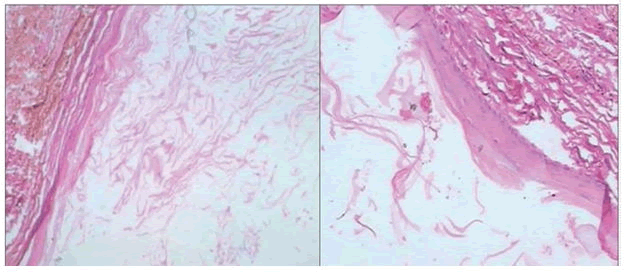
Histopathological examination confirmed the diagnosis of epidermoid cyst by presence of cystic lumen lined by stratified squamous epithelium with orthokeratin production. A total of five patients were reviewed: Three males and two females [5]. All the patients presented with asymptomatic postauricular extracranial cysts at the left postauricular side. Most patients sought medical intervention because of cosmetic reasons. There was no record of malignant transformation of the postauricular cysts. Complete surgical excision of the postauricular extracranial cysts was carried for all the lesions. No recurrence of the lesion was observed (Figure 3).
Discussion
Epidermoid and dermoid cysts of the head and neck region are uncommon; the usual site is commonly around the orbit. Developmental cyst(s) rarely occur at the postauricular site and this could explain why we had few cases during the study period.
Studies on the head and neck epidermoid and dermoid cysts irrespective of the sites showed that they could occur at any age. This study involves age group between 0-18 yrs. Studies have shown that in the postauricular region, epidermoid cyst are more common than dermoid cyst. Unilateral asymptomatic swelling is usually the main complaint of patients with postauricular cysts [6].
The cyst may rupture, be inflamed and be painful. However, in this present study, all the patients presented with asymptomatic, unilateral, painless and unruptured postauricular swellings. We routinely do an ultrasound for any postauricular cyst as the first line of radiological imaging and computed tomographic scanning is requested for any suspicious intracranial extension. All the patients in this study had postauricular extracranial cysts. The main reason for seeking medical intervention was for cosmetic reasons [7]. The need for early presentation and treatment cannot be overemphasized to forestall this risk, as malignant transformation, though rare, carries a poor prognosis. Recurrence of epidermoid and dermoid cysts may occur after incomplete excision. Hence, there is a need for reasonable follow-up after surgical excision to detection and treat the cyst recurrence.
Conclusion
In conclusion, postauricular epidermoid cysts are rare in the head and neck region. Asymptomatic nature of this condition most probably contributes to the patient’s delay in seeking early medical intervention. The need to create medical awareness for any disease conditions especially for asymptomatic conditions such as cutaneous cysts cannot be overemphasized to prevent malignant transformation, which carries a poor prognosis.
References
- Jyothi SR, et al. Epidermoid cysts of head and neck. Journal of Evolution of Medical and Dental Sciences, Vol. 2, No. 39, 2013, pp. 7533-7536.
[Crossref]
- Cho Y and Lee DH. Clinical characteristic of idiopathic epidermoid and dermoid cyst of the ear. Journal of Audiology and Otology, Vol. 21, No. 2, 2017, pp. 77-80.
[Crossref] [Google Scholar] [PubMed]
- Dutta M, et al. Epidermoid cyst in head and neck: Our experiences, with review of literature. Indian Journal of Otolaryngology and Head and Neck Surgery, Vol. 65, No. 1, 2013, pp. 14-21.
[Crossref] [Google Scholar] [PubMed]
- de Souza BA, et al. A rare case of dermoid cyst behind the ear. Plastic and Reconstructive Surgery, Vol. 112, No. 7, 2003, pp. 1972.
[Crossref] [Google Scholar] [PubMed]
- Al-Khateeb TH, et al. Cutaneous cysts of the head and neck. Journal of Oral and Maxillofacial Surgery, Vol. 67, No. 1, 2009, pp. 52-57.
[Crossref] [Google Scholar] [PubMed]
- Dive AM, et al. Epidermoid cyst of the outer ear: A case report and review of literature. Indian Journal of Otology, Vol. 18, No. 1, 2012, pp. 34.
- Awasthi N. Postauricular dermoid cyst: An unusual presentation. International Journal of Health and Allied Sciences, Vol. 6, No. 2, 2017, pp. 121-122.


