Research - International Journal of Medical Research & Health Sciences ( 2021) Volume 10, Issue 2
Preparation of Bio-Bandage from Human Platelet Lysate Admixed with Sericin Polymer for Efficient Wound Healing
Veeresh Nandikolmath1, Lakshmi Kanth RN2, Sandeep Kumar Padhy2, Manas Ranjan Padhy2 and Sharangouda J Patil1*2Department of Biotechnology, Ramaiah College of Arts, Science and Commerce, Bengaluru, Karnataka, India
Sharangouda J Patil, Stroma Biotechnologies Private Limited, Bengaluru, Karnataka, India, Email: shajapatil@gmail.com
Received: 30-Dec-2020 Accepted Date: Feb 11, 2021 ; Published: 18-Feb-2021
Abstract
Objectives: A bandage is typically a material that is used to support a dressing, splint, or other medical devices, or it can be used to provide mechanical support to part of the body. The dressing is the component that is applied directly to the wound and is responsible for promoting wound healing. After a skin lesion, the healing process requires the recruitment and activity of different cell types, such as native immune response cells, endothelial progenitor cells, keratinocytes, and fibroblasts.
Materials and methods: Platelet concentrate bag was obtained by apheresis unit (3 days before expiration) from Food and Drug Administration (FDA) regulated blood bank, shipped to the lab at 4°C-8°C temperature using an ice pack, and was placed in storage at -80°C until use. Silk cocoons (Bombyx mori) were collected from the Ram Nagar Silk Cocoon market in well-sterilized plastic bags at ambient temperature and shipped to the lab and stored at room temperature till the cocoons were processed.
Results: The platelet count was (18.5 ± 2.3) × 109 cells/ml. The pH of human Platelet Lysate (hPL) and sericin were checked by digital pH meter and were found 6.8 ± 0.1at 27°C and 6.9 ± 0.1 at 26.5°C respectively. Similarly, hPL and sericin were tested for sterility by the spread plate method. The moisture content of the hPL powder was analyzed by the Karl Fischer method and it was found 1.842% w/w.
Conclusion: Sericin was successfully isolated by the non-chemical method and exhibited good gelling property, became soft and hydrophilic compared to the chemical method, suggesting that sericin may be more suitable for application as a scaffold (wound dressing). Thus our study shows this admixture material is a suitable biological material for many applications in wound management
Keywords
Sericin, Human platelet lysate, Wound healing, Platelet Rich-Plasma (PRP), Bio-Bandage, Scaffold
Introduction
A bandage is typically a material that is used to support a dressing, splint, or other medical devices or it can be used to provide mechanical support to part of the body. The dressing is the component that is applied directly to the wound and is responsible for promoting wound healing. Depending on the composition, the dressing can function in a multitude of ways and while traditionally they would have been used to stop bleeds, absorb exudates and minimize contamination, they can comprise of an intricate composite with constituents designed to ease pain, combat infection, and actively promote healing through directly influencing the wound dynamics. It is impossible to pinpoint the first use of a bandage but given the discovery of dyed flax fibers and textiles close to linen that date back some 36,000 years [1]. Even the very earliest of societies likely could manufacture and employ some form of bandage. The needs for advanced wound dressing are traditionally used to protect the wound from contamination [2]. The use of topical bioactive agents in the form of solutions, creams, and ointments for drug delivery to the wound is not very effective as they rapidly absorb fluid and in the process lose their rheological characteristics to become mobile [3]. For this reason, the use of solid wound dressings is preferred in the case of exudative wounds as they provide better exudate management and prolonged residence at the wound site.
The introduction of the next generation of biological bandages (wound dressings) could stimulate regeneration by the release of a well-proportioned combination of growth factors [4]. An ideal wound dressing should satisfy some conditions such as good biocompatibility, the capacity for blocking dust and bacteria from entering the wound, good permeability, and the ability to promote the efficient formation of epithelial tissue. Silk consists of fibroin; sericin and silk sericin is a type of natural polymer fiber protein extracted from silk that has been widely used in biomedical purposes owing to its unique physical, mechanical and biological properties, such as good elasticity, low molecular weight, good biocompatibility and lack of pro-inflammatory effects [5,6]. Silk sericin contains peptides and can promote the differentiation and proliferation of human skin fibroblasts [7]. Additionally, the nano-fibrous scaffolds could improve the adhesion and proliferation of human skin fibroblasts. Although all of these materials improve wound healing, they have several drawbacks, including complex preparation of protocols and single structures. Furthermore, the extraction of silk sericin from natural cocoons can disrupt the structure of the cocoons to some extent. Natural cocoons are known to show good permeability and may have several benefits.
For example, the good permeability will prevent the area surrounding the wound from becoming damp, which would promote wound healing. Human Plasma Lysate (hPL) is a type of plasma containing highly concentrated platelets, produced by gradient density centrifugation of whole blood. After lysing, platelets can release numerous growth factors such as Basic Fibroblast Growth Factor (BFGF), Vascular Endothelial Growth Factor (VEGF), Epidermal Growth Factor (EGF), which plays an important role in the wound healing process [8,9]. Some reports have demonstrated that Plate Rich-Plasma (PRP) can promote cell proliferation and accelerate the process of wound healing [10]. PRP could not only accelerate wound healing but also reduce the risk of serious wound infections [11,12]. However, the high sensitivity and short half-lives of these factors have limited their applicability and made their bioactivities unstable [13]. This problem may be overcome by combining hPL with porous materials allowing for the sustained release of components and therefore prolonging the active time of the material. Additionally, for better results we can combine the Cocoon Scaffolds (CCSs) with hPL wherein CCSs could act as storage units for growth factors (BFGF, VEGF, and EGF) contained in hPL. Thus, showing CCSs admixed with hPL could have new biomedical applications in wound healing.
The human body responds to injury by attempting to heal itself. Regenerative medicine accelerates this natural healing process by harnessing advances in physiology, pathology, and pharmacology, to restore the health and function of cells, tissues, and organs compromised by disease, aging, or congenital defects [14]. After a skin lesion, this healing process requires the recruitment and activity of different cell types, such as native immune response cells, endothelial progenitor cells, keratinocytes, and fibroblasts. As in this context, a therapeutic approach to repair or regenerate the tissue is needed, the traditionally employed dressing materials used to cover and protect wounds have been substituted with modern wound dressings, also conceived to enhance the re-epithelialization, collagen synthesis, and angiogenesis and/or to prevent infections [15,16]. Among them, human blood materials and some natural compounds have been tested. In particular, Platelet Lysate (PL) has emerged as an effective medium supplement for the expansion of various cell types; other autologous or allogeneic human blood-derived materials, including human serum and plasma, have been tested, but they were not as promising as PL [17]. Recently, also Silk Sericin (SS), a natural protein isolated from cocoons, has been exploited in the last decades due to its remarkable properties [18]. Biomaterials employed in tissue engineering have a pivotal role in the regenerative process: they do not act only as “filler” providing structural support for cell accommodation and attachment, but they also guide and sustain new tissue formation and maturation [19].
Human Platelet Lysate
Human Platelet Lysate (hPL) is the product obtained by platelet lysis. Platelets are corpuscular blood elements, which play an essential role in the regulation of hemostasis and clot formation during bleeding, but also in the wound healing and tissue regeneration processes. In case of tissue damage, the adhesion molecules exposed by the damaged endothelium (such as the Von Wille brand factor, collagen, fibronectin, and laminin) activate platelets that release growth factors previously stored in their α-granules. This process triggers the tissue regeneration mechanism [20]. The same growth factors released in physiological conditions are useful for the production of cell culture supplements. The use of hPL for wound healing has several advantages: first of all, has no xenogeneic proteins, so it is free of that immunological risks. Then, in Europe, after the approval of European directive 2001/83/CE, blood-derived products (and among them hPL), are considered as medical products and they must be manufactured in compliance with the principles of Good Manufacturing Practice (GMP), to meet the requirements of quality, safety, and efficacy. In this way, the obtained product is standardized, traceable, and in the absence of viral, bacterial, or prion contaminations [21]. The hPL efficacy on the proliferation of different cell lines has been reported by different authors. However, an effective medium supplement should not only support the cell proliferation but even maintain the cell phenotype [22].
Platelet loaded into a silk fibroin-based scaffold, allowed a complete wound closure on a murine model within 6 days, while on the clinical practice, wound treated with PL gel recovered more quickly in all patients, leading to a reduction of the hospital stay and the related costs [23]. No adverse reactions were observed and all patients treated with platelet gel reported significant pain relief, thus bettering their quality of life [24]. Moreover, it has emerged that peptides or proteins, most likely belonging to the complement system, play a major role in the inhibition of Escherichia coli, Pseudomonas aeruginosa, Staphylococcus aureus, and Klebsiella pneumoniae [25]. Therefore, the inclusion of PL into scaffolds can lead to significant benefits not only in terms of regeneration of the damaged tissue but also to control and reduce the risk of infections [26].
Silk Sericin
Silk Sericin (SS) is a globular and water-soluble protein with a molecular weight of up to 200 kDa. Silk presents almost all characteristics desirable for biomedical applications, including biocompatibility, low toxicity, mechanical stability, and biodegradability [27]. Sericin has been widely used for the manufacturing of scaffolds for tissue engineering and regenerative medicine, exploiting its ability to improve cell adhesion and proliferation [28]. Nowadays, sericin emerged as an important biomaterial with several applications in different fields, including cosmetic, pharmaceutical, and delivery systems to administer drugs or cells [29,30]. Recently, Zhou, et al. characterized SS and SS-based nanoparticles in terms of antibacterial action against Gram-negative (E. coli) and Gram-positive (S. aureus) bacteria models [31]. Interestingly, these different biological activities are related to different SS molecular weight. The SS was added to the cell culture media for its ability to protect cells from oxidative stress, its anti-apoptotic properties, and also for its mitogen effect. SS can also be used to treat burn-wounds, reduce scars, and prevent infections [32]. Thus, SS can be employed, formulated into wound dressings or creams, to control wound infections. For example, in one clinical study, involving 29 patients with second-degree burn wounds, complete healing was obtained 5-7 days earlier after treatment with a cream containing SS concerning the control (silver zinc sulfadiazine cream) [33]. Recently, the antimicrobial effect of SS for wound healing has been improved by combining sericin based-scaffolds with silver nanoparticles [34]. Due to the novelty of PRP is been found to be effective in several case-control studies in addition to several non-controlled clinical trials. PRP has been suggested for their bacterial growth inhibition [35]. PRP treatment improves the healing capacities of human Multipotent Adipose-Derived Stem (hMADS) cells following their engraftment into mouse wounds [36]. To determine whether PRP treatment could improve the ability, hMADS cells and PRP were simultaneously delivered into mouse wounds. PRP treatment exerts cellular protective functions and counteracts metabolism dysfunctions induced by H2O2 injury in hMADS cells by restoring their mitochondrial respiration. PRP has recently been shown to reduce the incidence of sternal infections after cardiac surgery. PRP has also found popular and effective applications in sports and sports medicine in mending injured ligaments and tendons without surgeries [37].
Materials and Methods
Procurement of Platelet Concentrate Bags
Platelet concentrate bag was obtained by apheresis unit (3 days before expiration) from FDA-regulated blood bank, shipped to the lab at 4°C-8°C temperature using an ice pack, and was placed in storage at -80°C until its use. Donors of the platelet concentrate unit were screened and tested by blood centers for viral markers (i.e., hepatitis B virus, hepatitis C virus, human immunodeficiency, and syphilis) by kit method (Bio-rad).
Preparation of Platelet Lysate
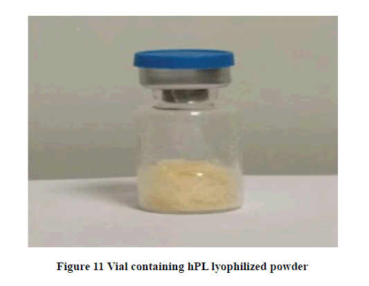
A research-scale lot of hPL was manufactured adopting the following method. Leuco-reduction of Platelet Concentrate (PC) bag was done by spinning unit of PC bag at 2000 rpm for 15 minutes at 20°C. A bag of 45 ml platelet concentrate was subjected to 2 cycles of freeze-thaw at 37°C to make platelet lysate. After the first round of freeze-thaw method, around 60%-70% of membrane ruptures, and for a better result the second round of freeze-thawing was done which gives results of membrane ruptures around 90%. After thawing, still, some membrane fragments were left, which were removed using cell strainers (Millipore, pore size 100 micron). Finally, strained platelet lysate was collected and filtered by filtration using a Sterile Filtration unit (Tarson, Germany) for fungus/bacteria. Then the filtrate product is collected in a sterile container. 1 ml of sterile product is transferred into each 2 ml capacity sterile glass vials with partial closure by rubber stopper under GMP protocol. Filled vials were transferred into a lyophilizer chamber, where freezing and vacuum drying was carried out till the product is completely made into powder. The rubber stoppers were closed once the procedure was completed and the vials were sent for aluminium crimping aseptically. The entire lyophilization process last for 18½ hrs and the vials were ready. Each vial contained (80-100) mg of lyophilized powder and was stored at room temperature until use.
Lyophilization Cycle
The filled vials were capped loosely and loaded on to the lyophilizer chamber. Vials were subjected for freezing/ precooling for 45 min at -40°C followed by primary drying for 15 hrs at -20°C (sublimation) under vacuum. Then secondary drying for 3 hrs (desorption).
Preparation of Nutrient Agar Media
2.8 g of nutrient agar (Hi-Media, M001-100G) was suspended in 250 ml of the conical flask containing 100 ml of distilled water. The conical flask was boiled to dissolve the medium completely and was sterilized by autoclaving at 15 lbs pressure (121°C) for 15 minutes. Then the conical flask was kept for cooling at room temperature (27°C). After cooling, the nutrient agar was poured into the plate.
Sterility Test
The microbial sterility of the hPL powder was tested by reconstituting 100 mg powder in 1000 μl WFI. From this, 50 μl was gently distributed by spread plate method over nutrient agar (Hi-Media, M001-100G) to reveal the bacterial or fungal (molds and yeast) contamination respectively. After seeding, the nutrient agar plates were maintained at 32.5°C for 5 days. Plates were checked for the presence of fungal and bacterial colonies.
Procurement of Silk Cocoons
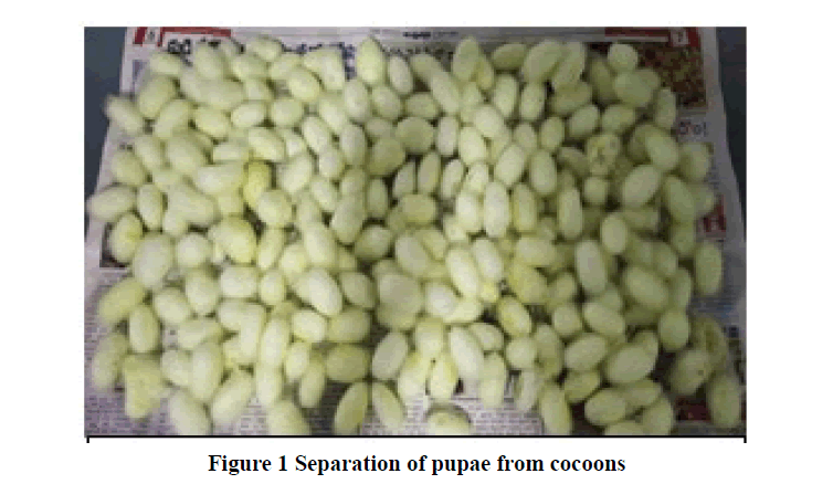
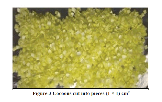
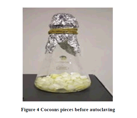
Silk cocoons (Bombyx mori) were collected from the Ram Nagar Silk Cocoon market in well-sterilized plastic bags at ambient temperature and shipped to the lab and stored at room temperature till the cocoons were processed. 1000 g of silk cocoons (Figure 1) were weighed in a weighing machine (Citizen Scale, CY 220C) and cleaned by handpicking (Figure 2). Cleaning by handpicking method removes small and large dirt particles properly. After cleaning, the cocoons were cut into small pieces of dimension (1 × 1) cm (Figure 3) and washed under running tap water for 5 minutes. After washing, the pieces were spread on aluminium foil for drying at room temperature for 24 hours. Then again the pieces were immersed in cold water for 10 minutes followed by drying again at room temperature for 24 hrs. After drying, pieces were weighed again and transferred into a conical flask for sterilization. 2 g and 5 g of cocoon pieces were weighed and transferred to a 250 ml conical flask containing 50 ml of double-distilled water separately, flasks were plugged with cotton. Then the conical flasks were sterilized by wet heat method (autoclaving) (Figure 4) at 121°C for 20 minutes in 15 lbs pressure. After sterilizing, the sericin extract of the two conical flasks was collected in two separate 15 ml sterile disposable centrifuge tubes and stored at 4°C until their use. The physical properties like pH and sterility were checked by digital pH meter and nutrient agar spread plate method respectively. After incubating the nutrient agar plates were maintained at 32.5°C for 5 days. Plates were checked for the presence of fungal and bacterial colonies.
Statistical Analysis
All the experiments were carried out in triplicates and were expressed as mean difference of standard deviation ± standard error. The data were statistically analyzed using Microsoft Office Excel 2007.
Results
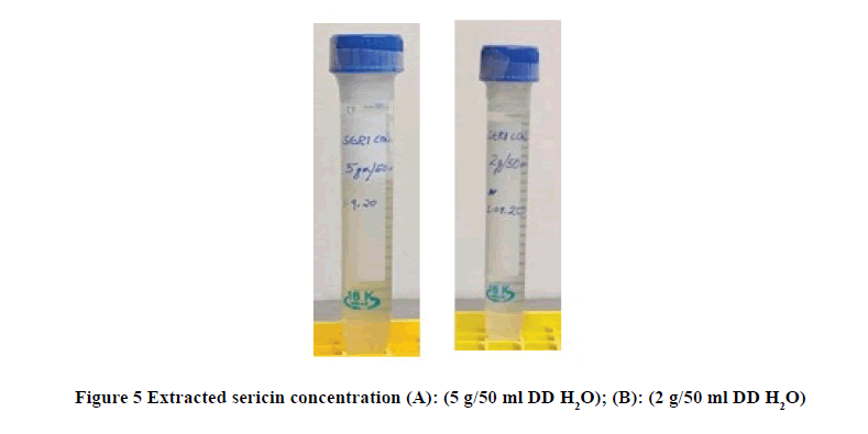
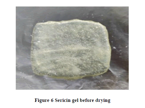
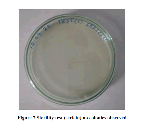
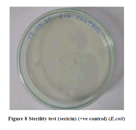
Sericin is a natural biopolymer produced by silkworm (Bombyx mori), which is bound with two fibroin filaments together like a thread of silk to by the cocoon. In the present study, sericin was isolated from the silk cocoon. Silk cocoons were sterilized by wet heat method and checked for sterility and pH. The sericin volume obtained from flask A (5 g in 50 ml of double-distilled water) is 8.2 ml (Figure 5). The volume of sericin obtained from flask B (2 g in 50 ml of double-distilled water) is 17 ml (Figure 5). Further sericin gel is prepared from flask A (5 g in 50 ml of doubledistilled water) and cast on a sterile aluminium sheet and kept for drying at 37°C (Figure 6). The sterility of extracted sericin of both the volume was checked by the spread plate method and was found sterile (Table 1, Figures 7, 8, and 9). The pH for isolated sericin of flask A was checked by a digital pH meter and was found to be 6.9 ± 0.1 at 26.5°C (Table 2). The pH for isolated sericin of flask B was checked and found to be 6.7 at 26.5°C.
Table 1 Sterility test sericin
| Temp. | Test type | Incubation time | Observation |
|---|---|---|---|
| (in days) | |||
| 32.5°C | Negative Control | 5 | No Colonies |
| 32.5°C | Positive Control | 5 | 3 Colonies |
| 32.5°C | Test 1 | 5 | No Colonies |
| 32.5°C | Test 2 | 5 | No Colonies |
| 32.5°C | Test 3 | 5 | No Colonies |
Table 2 pH test sericin (at 26.5°C)
| Sericin | pH |
|---|---|
| 5gm (8.2 ml) | 6.9 ± 0.1 |
| 2gm (17 ml) | 6.7 ± 0.1 |
| D.D/w | 7 |
Data are presented as means ± SD
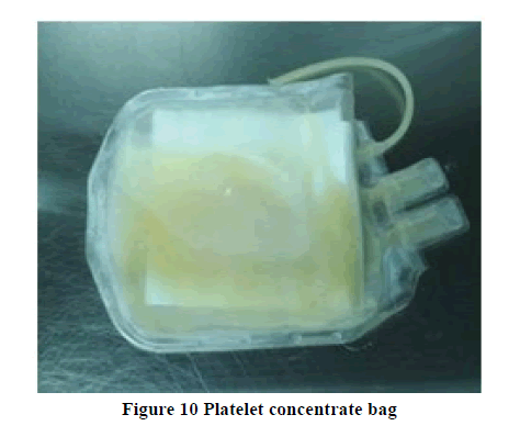
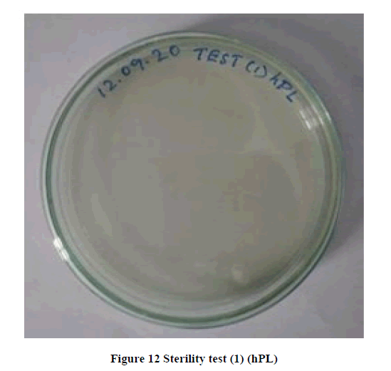
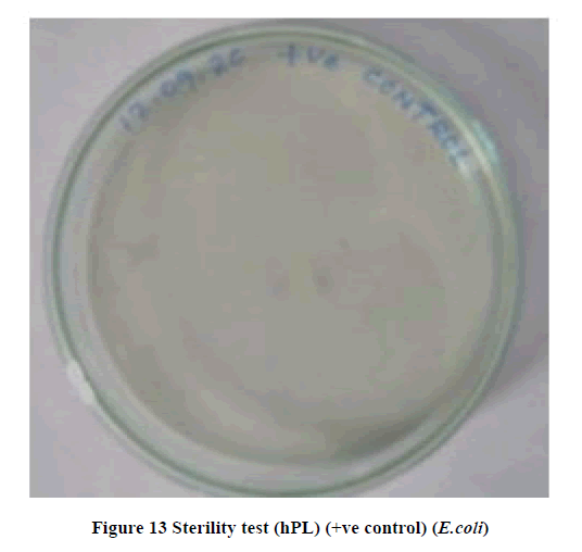
In the present study, PC bag (Figure 10) content was measured and found 45 ml and it was screened for infectious diseases by rapid kit method (Bio-Rad) and was found negative for Syphilis, Human Immunodeficiency Virus, Hepatitis C Virus and Hepatitis B Virus respectively. The platelet count was (18.5 ± 2.3) × 109 cells/ml. The volume of hPL obtained after separation of membrane fragments by centrifugation and filtration was 40.5 ml. After centrifugation, 1 ml of hPL was dispensed into each 2 ml capacity glass vial and was placed in a lyophilizer chamber for 18½ hrs. (80-100) mg hPL was obtained in each vial (Figure 11). The sterility of hPL powder was checked by the nutrient agar spread plate method and was found sterile (Table 3 and Figures 12, 13, and 14). The pH of hPL was checked in a digital pH meter and was found 6.8 ± 0.1. The moisture content of the lyophilized powder was analyzed by the Karl Fischer method and it was 1.842% w/w (Table 4).
Table 3 Sterility test hPL
| Temp. | Test type | Incubation time | Observation |
|---|---|---|---|
| (in days) | |||
| 32.5°C | Negative Control | 5 | No Colonies |
| 32.5°C | Positive Control | 5 | 3 Colonies |
| 32.5°C | Test 1 | 5 | No Colonies |
| 32.5°C | Test 2 | 5 | No Colonies |
| 32.5°C | Test 3 | 5 | No Colonies |
Table 4 pH test hPL (at 27°C)
| hPL | pH |
|---|---|
| 100 mg in 1 ml | 6.8 ± 0.1 |
Data are presented as means ± SD
Discussion
The biological properties of sericin application depend on the culture medium, cryopreservation, used in drug delivery system, demonstrating the effective utilization in tissue engineering and act as a novel biomaterial molecule. Due to the low digestibility of sericin molecule explored it the application of medical field for the biological activities, such as antitumor, antimicrobial, anti-inflammatory agent, anticoagulant, colon health support, improvement of constipation, and also protection in obesity by improved lipid plasma therapy [38]. Sterility of extracted sericin confirmed by spread plate method and pH also obtained from isolated sericin. These findings were similarly revealed with the Rocha, et al. to determine extraction, physicochemical, biological characterization, and their biopharmaceutical applications [39]. In human Platelet Lysate (hPL), 45 ml was found potential against infectious diseases by the rapid kit method and was found negative for Syphilis, HIV, HCV, and HBV respectively. Thus, it has evident with the study of Jantaruk, et al. as an application of oligopeptides obtained from sericin for the treatment of infectious diseases and tumors [40]. Using such biomolecule as a bio-bandage agent against such infectious organisms is a novel finding and it’s a challenge to the dermatologist and pathologist to combat the errors of inflammation, wound healing, and other infections from barriers. Padamwar and Pawar explored such protein for the applications in cosmetics and pharmaceuticals as a biological agent for wounds, bio-adhesive moisturizing, anti-wrinkle, and anti-aging [41]. It is due to their nano-fibrous structural mat will support by the dressing and constructing as tissue repair and regeneration by using biopolymer polyvinyl alcohol incorporated with silk sericin [42].
Conclusion
In conclusion, sericin was successfully isolated by a non-chemical method, exhibited good gelling property, become soft, hydrophilic compared with chemical methods, and suggesting that sericin may be more suitable for application as a scaffold (wound dressing). Similarly, hPL was prepared by lyophilization and tested for its physic-chemical properties. Further in vitro and in vivo investigations are required to study the significance of a judicious combination of sericin with hPL in the promotion of wound healing. Moreover, previous reports of in vivo (animal model) studies showed that CCSs + PRP could accelerate wound healing more effectively. Thus our study shows this admixture material is a suitable biological material for many applications in wound management.
Declerations
Acknowledgments
The authors would like to thank Director, Ram Nagar Silk Cocoon Market, Ramnagar, Karnataka, India for providing the cocoons for the studies and support in research.
Conflict of Interest
The authors declared no potential conflicts of interest with respect to the research, authorship, and/or publication of this article.
References
- Kvavadze, Eliso, et al. "30,000-year-old wild flax fibers." Science, Vol. 325, No. 5946, 2009, p. 1359.
- Sood, Aditya, Mark S. Granick, and Nancy L. Tomaselli. "Wound dressings and comparative effectiveness data." Advances in Wound Care, Vol. 3, No. 8, 2014, pp. 511-29.
- Boateng, Joshua S., et al. "Wound healing dressings and drug delivery systems: a review." Journal of Pharmaceutical Sciences, Vol. 97, No. 8, 2008, pp. 2892-923.
- Fife, Caroline E., and Marissa J. Carter. "Wound care outcomes and associated cost among patients treated in US outpatient wound centers: Data from the US wound registry." Wounds: A Compendium of Clinical Research and Practice, Vol. 24, No. 1, 2012, pp. 10-17.
- Kanokpanont, Sorada, et al. "An innovative bi-layered wound dressing made of silk and gelatin for accelerated wound healing." International Journal of Pharmaceutics, Vol. 436, No. 1-2, 2012, pp. 141-53.
- Kundu, Joydip, et al. "Silk fibroin/poly (vinyl alcohol) photocrosslinked hydrogels for delivery of macromolecular drugs." Acta Biomaterialia, Vol. 8, No. 5, 2012, pp. 1720-29.
- Yamada, Hiromi, et al. "Identification of fibroin-derived peptides enhancing the proliferation of cultured human skin fibroblasts." Biomaterials, Vol. 25, No. 3, 2004, pp. 467-72.
- Bacakova, Lucie, et al. "Nanofibrous scaffolds for skin tissue engineering and wound healing based on synthetic polymers." Applications of Nanobiotechnology, 2019, pp. 33-61.
- Alves, Rubina, and Ramon Grimalt. "A review of platelet-rich plasma: History, biology, mechanism of action, and classification." Skin Appendage Disorders, Vol. 4, No. 1, 2018, pp. 18-24.
- Chicharro-Alcántara, Deborah, et al. "Platelet rich plasma: New insights for cutaneous wound healing management." Journal of Functional Biomaterials, Vol. 9, No. 1, 2018, p. 10.
- Everts, Peter AM, et al. "Platelet-rich plasma and platelet gel: a review." The Journal of Extra-Corporeal Technology, Vol. 38, No. 2, 2006, pp. 174-87.
- Carter, Marissa J., Carelyn P. Fylling, and Laura KS Parnell. "Use of platelet rich plasma gel on wound healing: a systematic review and meta-analysis." Eplasty, Vol. 11, 2011, p. e38.
- Liu, Jiawei, et al. "Healing of skin wounds using a new cocoon scaffold loaded with platelet-rich or platelet-poor plasma." RSC Advances, Vol. 7, No. 11, 2017, pp. 6474-85.
- Chen, Fa-Ming, and Xiaohua Liu. "Advancing biomaterials of human origin for tissue engineering." Progress in Polymer Science, Vol. 53, 2016, pp. 86-168.
- Lee, Kangwon, Eduardo A. Silva, and David J. Mooney. "Growth factor delivery-based tissue engineering: general approaches and a review of recent developments." Journal of the Royal Society Interface, Vol. 8, No. 55, 2011, pp. 153-70.
- Dhivya, S., V. V. Padma, and E. Santhini. "Wound Dressings-A Review. Biomedicine (Taipei), 5, 22." 2015.
- Hemeda, Hatim, Bernd Giebel, and Wolfgang Wagner. "Evaluation of human platelet lysate versus fetal bovine serum for culture of mesenchymal stromal cells." Cytotherapy, Vol. 16, No. 2, 2014, pp. 170-80.
- Aramwit, Pornanong, et al. "The effect of sericin from various extraction methods on cell viability and collagen production." International Journal of Molecular Sciences, Vol. 11, No. 5, 2010, pp. 2200-11.
- Green, David W., et al. "Calcifying tissue regeneration via biomimetic materials chemistry." Journal of the Royal Society Interface, Vol. 11, No. 101, 2014, p. 20140537.
- Rumbaut, Rolando E., and Perumal Thiagarajan. "Platelet-vessel wall interactions in hemostasis and thrombosis." Synthesis Lectures on Integrated Systems Physiology: From Molecule to Function, Vol. 2, No. 1, 2010, pp. 1-75.
- AuBuchon, James P., et al. "Efficacy of apheresis platelets treated with riboflavin and ultraviolet light for pathogen reduction." Transfusion, Vol. 45, No. 8, 2005, pp. 1335-41.
- Strunk, Dirk, et al. "International Forum on GMP‐grade human platelet lysate for cell propagation." Vox Sanguinis, Vol. 113, No. 1, 2018, pp. e1-e25.
- Ma, Kun, et al. "Effects of nanofiber/stem cell composite on wound healing in acute full-thickness skin wounds." Tissue Engineering Part A, Vol. 17, No. 9-10, 2011, pp. 1413-24.
- Mazzucco, Laura, et al. "The use of autologous platelet gel to treat difficult‐to‐heal wounds: a pilot study." Transfusion, Vol. 44, No. 7, 2004, pp. 1013-18.
- Chen, Fa-Ming, et al. "A review on endogenous regenerative technology in periodontal regenerative medicine." Biomaterials, Vol. 31, No. 31, 2010, pp. 7892-927.
- Lee, T. Clive, and Peter Niederer, eds. "Basic engineering for medics and biologists: an ESEM primer." IOS Press, Vol. 152, 2010.
- Liu, Wenying, Stavros Thomopoulos, and Younan Xia. "Electrospun nanofibers for regenerative medicine." Advanced Healthcare Materials, Vol. 1, No. 1, 2012, pp. 10-25.
- Jones, Gemma L., et al. "Osteoblast: osteoclast co-cultures on silk fibroin, chitosan and PLLA films." Biomaterials, Vol. 30, No. 29, 2009, pp. 5376-84.
- Okazaki, Yukako, et al. "Consumption of a resistant protein, sericin, elevates fecal immunoglobulin A, mucins, and cecal organic acids in rats fed a high-fat diet." The Journal of Nutrition, Vol. 141, No. 11, 2011, pp. 1975-81.
- GAMO, TAKUMA. "Biochemical genetics and its application to the breeding of the silkworm." Japan Agriculture Research Quarterly, Vol. 16, No. 4, 1983, pp. 264-72.
- Zhou, Wenhao, et al. "Bioinspired and biomimetic AgNPs/gentamicin-embedded silk fibroin coatings for robust antibacterial and osteogenetic applications." ACS Applied Materials and Interfaces, Vol. 9, No. 31, 2017, pp. 25830-46.
- Rasulov, M. F., et al. "First experience in the use of bone marrow mesenchymal stem cells for the treatment of a patient with deep skin burns." Bulletin of Experimental Biology and Medicine, Vol. 139, No. 1, 2005, pp. 141-44.
- Robins, Elaine V. "Burn shock." Critical Care Nursing Clinics of North America, Vol. 2, No. 2, 1990, pp. 299-307.
- Vahabi, Khabat, G. Ali Mansoori, and Sedighe Karimi. "Biosynthesis of silver nanoparticles by fungus Trichoderma reesei (a route for large-scale production of AgNPs)." Insciences Journal, Vol. 1, No. 1, 2011, pp. 65-79.
- Marx, Robert E. "Platelet-Rich Plasma (PRP): what is PRP and what is not PRP?." Implant Dentistry, Vol. 10, No. 4, 2001, pp. 225-28.
- Hersant, Barbara, et al. "Platelet-rich plasma improves the wound healing potential of mesenchymal stem cells through paracrine and metabolism alterations." Stem Cells International, Vol. 2019, 2019.
- Mishra, Allan, et al. "Sports medicine applications of platelet rich plasma." Current Pharmaceutical Biotechnology, Vol. 13, No. 7, 2012, pp. 1185-95.
- Kunz, Regina Inês, et al. "Silkworm sericin: properties and biomedical applications." BioMed Research International, Vol. 2016, 2016.
- Rocha, Liliam KH, et al. "Sericin from Bombyx mori cocoons. Part I: Extraction and physicochemical-biological characterization for biopharmaceutical applications." Process Biochemistry, Vol. 61, 2017, pp. 163-77.
- Jantaruk, Pornpimon, et al. "Augmentation of natural killer cell activity in vitro and in vivo by sericin-derived oligopeptides." Journal of Applied Biomedicine, Vol. 13, No. 3, 2015, pp. 249-56.
- Padamwar, M. N., and A. P. Pawar. "Silk sericin and its applications: A review." Journal of Scientific Industrial Research, Vol. 63, No. 4, 2004, pp. 323-29.
- Gilotra, Sween, et al. "Potential of silk sericin based nanofibrous mats for wound dressing applications." Materials Science and Engineering: C, Vol. 90, 2018, pp. 420-32.

