Research - International Journal of Medical Research & Health Sciences ( 2020) Volume 9, Issue 9
Neonatal Sonographic Hip Screening for Developmental Dysplasia of Hips
Aeisha Begum1*, Salik Kashif2, Salma Ambreen1 and Laila Khan32Department of Orthopedic and Spine Surgery, Hayatabad Medical Complex, Peshawar, Pakistan
3Radiology Department, Lady Reading Hospital, Peshawar, Pakistan
Aeisha Begum, Radiology Department, Wah Medical College, Wah Cantonment, Pakistan, Tel: 091921714047, Email: aeishasalman@gmail.com
Received: 29-Jul-2020 Accepted Date: Sep 09, 2020 ; Published: 16-Sep-2020
Abstract
Ultrasound screening is recommended for newborn babies in many countries to rule out Developmental dysplasia of hip (DDH) as it is more sensitive and specific compared to clinical examination. The purpose of this study was to perform a preliminary assessment of neonatal hips in our region and highlight the magnitude of DDH as there is lack of local literature on this subject. We evaluated 132 hips newborns referred to our department using Graf static method of alpha and beta angle measurement and classification. The data collected was statistically correlated. Mean age was 1.5 days. There was no statistically significant difference between the mean alpha and beta angles of right and left side of hips or between male and female babies. The predominant hip type in both genders and on both sides was Graf Ia (75% of right and 69.9% left sided hips) followed by IIa. Although the vast majority of babies had normal or physiologically immature hips, a small number of children had abnormal hips requiring treatment. In order to diagnose the highest possible number of cases, we recommend further large scale studies targeting our population on the basis of which universal or selective screening programs for a wider clinical application could be based.
Keywords
Developmental dysplasia of Hip, Ultrasonography, Screening, Neonates
Introduction
Every newborn baby should be thoroughly examined and investigated for congenital anomalies including musculoskeletal anomalies. Some of the musculoskeletal congenital anomalies are obvious, such as clubfeet, constriction bands, limb length discrepancies, radial or ulnar club hand however Developmental dysplasia of hip (DDH) is a concealed anomaly. DDH encompasses a broad spectrum of developmental anomalies of the femur and acetabulum ranging from mild dysplasia to frank hip dislocation [1]. It is one of the important, common and potential preventable cause of disability with great socioeconomic implications [2]. As the condition is painless and the baby moves the hip joints quite well, it can frequently be overlooked. Delay in diagnosis and thus treatment results in extensive surgery with potential complications [3] and unrecognized and untreated cases can lead to premature osteoarthritis [1]. Risk factors [4,5] for DDH include positive family history, first pregnancy, breech presentation, female gender, oligohydramnios, limited hip abduction, talipes, swaddling and large birth size [6]. Female sex is the only isolated risk factor with a PLR (positive likelihood ratio) predictive of DDH [7]. In most cases the diagnosis of hip dislocation can be made by clinical examination of the newborn, but it is impossible to diagnose hip dysplasia (in which the hip is reduced but the acetabulum is shallow or underdeveloped) by clinical examination, additionally there may be a false positive click (e.g. snapping hip) on clinical examination. Hip dysplasia can easily be diagnosed by Ultrasound evaluation of the newborn’s hip by an experienced sonologist [8] a technique introduced initially in the 1980s [9,10]. A 15 year study found that ultrasound outperforms clinical examination, with a positive predictive value of 49% as compared to 24% with clinical examination [11]. In most of the developed countries ultrasound of the hip is performed in every single newborn, regardless of any risk factors [12], however the appropriate utilization of ultrasound screening for DDH is still controversial, with some countries and studies advocating universal screening, while others stressing selective screening (babies having two or more risk factors) [13,14]. The impact of ultrasound on diagnostic thinking and decision making of suspected DDH was 52% in a study [15].
High resolution ultrasound evaluates the relative position of femur and acetabulum. Alpha and beta angles are measured and hips classified according to the most widely used Graf method. Alpha angle determines the sonographic hip type and is formed between the straight lateral edge of ilium and bony acetabular margin. Beta angle is formed between straight lateral edge of ilium and fibrocartilaginous labrum, it determines the sonographic subtype of hip [1,13].
Ultrasound is readily available in almost all areas of the country and is an invaluable tool for diagnosis, management and surveillance of treatment [13]. Although, calculation of alpha and beta angles requires some expertise, but with minimal training, it can be easily learned and practiced. The inter-observer reliability of calculation of alpha and beta angles is very good [16,17]. Some radiologists use other methods [18] and dynamic assessment which evaluates the stability of the hip by observing the movement of the femoral head in and out of the acetabulum.
Our country does not have any screening program for neonatal hip assessment and there is lack of local literature on this subject. To the best of our knowledge, no local study addressing the incidence, presentation, diagnosis and management of DDH has been found in the local literature. The purpose of this study was to perform a preliminary assessment of neonatal hips in our region and highlight the magnitude of the DDH.
Materials and Methods
This study was performed in the Radiology department of tertiary care, POF hospital Wah cantt. Approval was taken from the ethical committee of hospital. Stable, new born babies were referred to our department for sonographic screening for developmental dysplasia of hips. All babies regardless of gender, period of gestation or mode of delivery referred to us with in the first week of their life were included in the study. Static hip assessment of both hips was done using 7.5 MHz high resolution linear probe with the baby lying on its side or lateral position with the hip slightly flexed. Scanning was done with a high frequency linear probe and the focus set at the acetabular edge. Alpha and beta angles for right and left hip were measured and documented by Graf method. These measurements were used to detect the presence of hip dysplasia and to classify it into different types using the most widely accepted Graf’s classification. Syndromic babies were excluded from the study.
Descriptive statistics were calculated for the alpha and beta angle of the right and left side hips of new born babies as well as gender-wise. Paired t-test was used to test the significance of differences between alpha and beta angles of the left and right hips. Independent samples t-tests was used to test the differences of angles of male and female babies and babies of age group 1 (less than 24 hour of age) and age group 2 (1 to 7 days age). Pearson correlations were calculated among right and left side, alpha and beta angles of all, female and male children. Trend of the relationship between alpha and beta angles were presented graphically. Numbers of children were calculated for the different groups according to Graf classification of hip developmental dysplasia.
Results
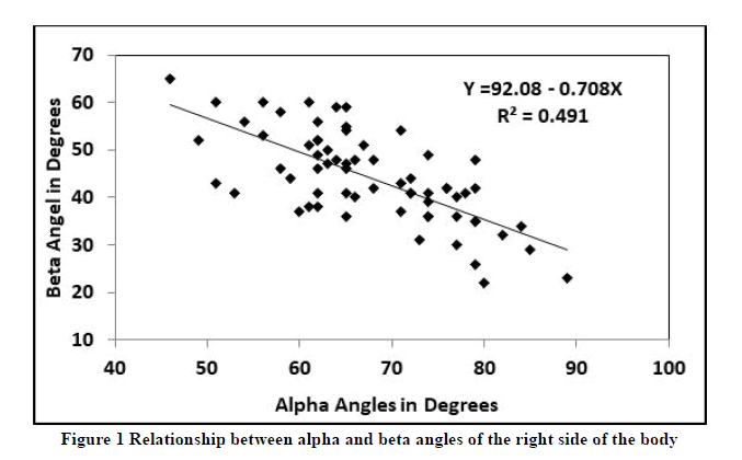
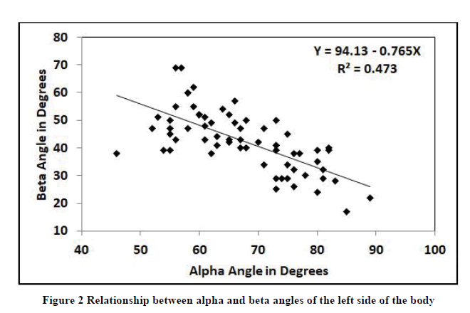
p>In this study alpha and beta angles of 132 hips of a sample of 66 babies were evaluated. About 64 per cent of the babies were less than 24 hours old at the time of scan, the rest of the babies were 1 to 7 days old; gender-wise there were 38 male and 28 female babies in the sample for the study. Sixty one babies were full term, two premature, and three post term; mode of delivery data showed that 37 of the babies were delivered by caesarean mode while 29 babies were delivered normal.The α angles ranged from 46 to 89°of both left and right sides with a mean of 67.6 ± 9.4° for the right side hip and 67.2 ± 9.7° for the left side hip (Table 1). The β angle ranged from 22 to 65°for the right sides with a mean of 44.2 ± 9.5°and it ranged from 17 to 69°with a mean of 42.7 ± 10.8°for the left side hip. Differences between mean α and β angles of the two sides was not significant based on paired t-test (Table 2) considering female, male and all babies. Differences between mean α and was β angles of male and female babies and between the two age groups was also not significant based on independent sample t-test (Table 3). The α and β angles the left hip as well as the right hip were highly negatively correlated (Table 4); the R2 of trend line shown in Figure 1 reveals that the regression model for β angle explain 49 of the variation in the alpha values for the right hip and the R2 of trend line shown in Figure 2 reveal that the regression model for the β angle explain 47 of the variation in alpha values for the left hip.
| Statistics | Right Side Angles in Degrees | Left Side Angles in Degrees | ||
|---|---|---|---|---|
| Alpha | Beta | Alpha | Beta | |
| All 66 children in the sample | ||||
| Average | 67.6 | 44.2 | 67.2 | 42.7 |
| Minimum | 46 | 22 | 46 | 17 |
| Maximum | 89 | 65 | 89 | 69 |
| Range | 43 | 43 | 43 | 52 |
| SD | 9.4 | 9.5 | 9.7 | 10.8 |
| Median | 65.5 | 43 | 66.5 | 42.5 |
| Female 28 children in the sample | ||||
| Average | 68.1 | 43.9 | 67.9 | 43.6 |
| Minimum | 46 | 23 | 46 | 28 |
| Maximum | 89 | 65 | 83 | 62 |
| Range | 43 | 42 | 37 | 34 |
| SD | 10.4 | 9.6 | 9.5 | 8.9 |
| Median | 71 | 44 | 67 | 43.5 |
| Male 38 children in the sample | ||||
| Average | 67.2 | 44.5 | 66.7 | 42 |
| Minimum | 49 | 22 | 52 | 17 |
| Maximum | 85 | 60 | 89 | 69 |
| Range | 36 | 38 | 37 | 52 |
| SD | 8.6 | 9.5 | 10 | 12.1 |
| Median | 65 | 42.5 | 64.5 | 41 |
| Categories | Alpha Angle in Degrees | Beta Angle in Degrees | |||||
|---|---|---|---|---|---|---|---|
| Gender | Side | Mean | SD | p-value | Mean | SD | p-value |
| Both | Left | 67.2 | 9.7 | 0.8301 | 42.7 | 10.8 | 0.3543 |
| Right | 67.6 | 9.4 | 44.2 | 9.5 | |||
| Female | Left | 67.9 | 10.4 | 0.9244 | 43.6 | 8.9 | 0.9073 |
| Right | 68.1 | 9.5 | 43.9 | 9.6 | |||
| Male | Left | 66.7 | 10 | 0.8475 | 42 | 12.1 | 0.2379 |
| Right | 67.2 | 8.6 | 44.5 | 9.5 | |||
| Categories | Alpha Angle in Degrees | Beta Angle in Degrees | |||||
|---|---|---|---|---|---|---|---|
| Mean | SD | p-value | Mean | SD | p-value | ||
| Female | Right Side | 68.1 | 10.4 | 0.6762 | 43.9 | 9.6 | 0.7959 |
| Male | 67.2 | 8.6 | 44.5 | 9.5 | |||
| Female | Left Side | 67.9 | 9.5 | 0.6271 | 43.6 | 8.9 | 0.5556 |
| Male | 66.7 | 10 | 42 | 12.1 | |||
| Age group 1 | Right Side | 66.4 | 9.6 | 0.1996 | 44.8 | 9.3 | 0.5365 |
| Age group 2 | 69.5 | 7.8 | 43.2 | 9.9 | |||
| Age group 1 | Left Side | 67.6 | 10.1 | 0.6812 | 42 | 10.7 | 0.5335 |
| Age group 2 | 66.6 | 9.3 | 43.8 | 11.2 | |||
| Right Alpha | Right Beta | Left Alpha | Left Beta | |
|---|---|---|---|---|
| All 66 Children † | ||||
| Right Alpha | 1 | -0.7010** | 0.1343 ns | 0.0115 ns |
| Right Beta | 1 | -0.0728 ns | 0.1432 ns | |
| Left Alpha | 1 | -0.6882** | ||
| Left Beta | 1 | |||
| Female 28 Children | ||||
| Right Alpha | 1 | -0.7677** | 0.2940 * | -0.1939 ns |
| Right Beta | 1 | -0.2476 * | 0.2598* | |
| Left Alpha | 1 | -0.6009** | ||
| Left Beta | 1 | |||
| Male 38 Children | ||||
| Right Alpha | 1 | -0.6468** | -0.0020 ns | 0.1385 ns |
| Right Beta | 1 | 0.0518 ns | 0.0870 ns | |
| Left Alpha | 1 | -0.7518 ** | ||
| Left Beta | 1 | |||
† ns not significant; *significant at the 5% level of probability; **significant at the 1% level of probability
Table 5 shows distribution of hip types by Graf classification according to sides i.e. right & left. Graf type Ia hips (normal and mature) were by far the most common constituting 72.45% of all hips with 75% for right hip and 69.9 for left hip. Graf type IIa (physiologically immature) hips were the second largest group accounting for 18.5% of the total hips. These two types are considered normal for new born babies, however Graf type IIa require a follow up ultrasound study at three months of age to see for the proper development of the hip. 2.25% hips fell in Graf IIc category which requires active management and treatment.
| Classification of Hip Developmental Dysplasia | Right Side | Left Side | Total | ||||
|---|---|---|---|---|---|---|---|
| Number | Percentage (%) | Number | Percentage (%) | Percentage (%) | |||
| Ia | Alpha ≥ 60° | Beta ≤ 55° | 50 | 75% | 47 | 69.9% | 72.45% |
| Ib | Alpha ≥ 60° | Beta >55° | 5 | 7.5% | 2 | 3% | 5.2% |
| IIa+ | α 50-59° | Beta >550 | 9 | 13.3% | 16 | 23.8% | 18.55% |
| IIc | α 43-49° | Beta <77° | 2 | 3% | 1 | 1.5% | 2.25% |
Table 6 shows distribution of hip types by Graf classification according to gender. 78.5% of male babies and 77.7% of female babies had normal mature hips (Graf type I) while 19.7% of male babies and 17.8% of female babies had physiologically immature hips (Graf type IIa). 1 (1.3%) male baby and 2 (3.5%) female babies had Graf IIc hips. We did not encounter Type 3 or type 4 hips in our study.
| Classification of Hip Developmental Dysplasia | Male Babies | Female Babies | Total | ||||
|---|---|---|---|---|---|---|---|
| Number | Percentage (%) | Number | Percentage (%) | Percentage (%) | |||
| Ia | Alpha ≥ 60° | Beta ≤ 55° | 55 | 72 | 43 | 76 | 74 |
| Ib | Alpha ≥ 60° | Beta >55° | 5 | 6.5 | 1 | 1.7 | 4.1 |
| IIa+ | α 50-59° | Beta >550 | 15 | 19.7 | 10 | 17.8 | 18.75 |
| IIc | α 43-49° | Beta <77° | 1 | 1.3 | 2 | 3.5 | 2.4 |
Discussion
Ultrasound of the newborn hip is now an established technique to confirm or refute the diagnosis of DDH. It not only diagnoses the dislocation but can also confirm and calculate the dysplastic acetabulum. In our study of 132 hips, 77.2% of babies had normal hips, 18.5% babies had immature hips and 3.75% babies had abnormal hips requiring treatment. There was no statistical significant difference between the alpha angles of the right hip (67.7) and that of the left hip (67.24) although in literature the left hip is more commonly involved [5] than the right hip in case of DDH, so theoretically the alpha angle should be lower on the left side as compared to the right side.
Jacobino, et al. [19] studied hips of 222 neonates with mean age of 5 days. By far the most common hip type in both genders and on both sides was Graf type Ia, with 78.4% of the right hips and 72% of left hips falling in this category. These results are comparable to our results with 75% of right hips and 69% of left hips identified as Graf Ia in our study. Their results showed that Mean α angle values were higher in males as well being higher for right side. In female babies, the mean α angle was 60.45°, whereas the mean β angle was 51.61° while in the male babies, the mean α angle was 62.8°, whereas the mean β angle was50.8°. In our series, the mean α angle in females was 68°, whereas the mean β angle was 43.75°. In male babies, the mean α angle was 66.95°, whereas the mean β angle was 43.25°. Our study found no statistically significant difference between the right and left sides or between both genders. The alpha angle measured in our study is much higher as compared to theirs and if we calculate the difference between our findings, in females the average alpha angle is 7.50 higher in our population and the average beta angle is 7.80 lower. In males the average alpha angle is 4.20 higher and the average beta angle is 7.50 lower. This indicates that our population have better developed acetabulae as compared to Brazilian population.
Daniel, [20] conducted hip ultrasound screening using the Graf static method in 65 neonates with mean age of 12 weeks. They reported that the mean alpha angles in female babies were 630 for the right hip and 640 for the left; whereas in the male babies, the mean alpha angles were 64.60 for the right hip and 630 for the left. The mean beta angles were 440 for the right hip and 450 for the left in females; while the mean beta angles were 470 for the right hip as well as the left hip in males. Our results show higher mean alpha angles for both male and female neonates. However similar to our results they also concluded that differences in both mean alpha and beta angles between males and females and between right and left sides were not statistically significant. Type I hip types were the predominant types on both sides and both genders in their study as well as our study.
Lussier, et al. [21] screened neonates for hip dysplasia, in their study they found out that 86.6% of the newborns screened within 28 days of birth had Graf type 1 hips, while 77.2% of our hips were Graf type 1. However they recommended to screen the hips by ultrasonography at 4 weeks of age, as it improves specificity with reduced false positives and follow-up visits and also reducing overtreatment, but this method of delayed ultrasonography is not suitable in our country because we do not have any screening program and many of them will lose follow-up resulting in neglect of the condition. Rawlings stated that the exact timing of ultrasound does not make much difference [22].
A study done on 8,356 Mongolian neonates with median age of 1 day at the time of ultrasound examination found that 89.0% had mature hips (Graf type I), 10.3% had physiologically immature hips (Graf type IIa), 0.2% had dysplastic, centered hips (Graf IIc), 0.4% dysplastic, decentered hips (Graf D), 0.08% subluxated hips (Graf 3), and 0.02% had dislocated hips (Graf type 4 hips) [23]. Their results differ from our results as in our study 77.2% had Graf type I hips, 18.5% had Graf IIa and 3.75% had Graf IIc hips.The most likely reason for this difference is our small sample size which does not represent the true picture of the incidence and spectrum of DDH.
Radiographs are not diagnostic early in life as the femoral head is not ossified till about 4 to 6 months of age thus not visible on X-rays and they also have the inherent risks of ionizing radiation. An alternate method is the use of arthrography, which is a very good technique to assess the hip joint, it clearly shows the femoral head, the shape of the acetabulum, status of the limbus and gives some idea about psoas constriction. But this is a very invasive method, requires special expertise and general anesthesia for it to be performed. Conventional arthrography is now being replaced by Magnetic Resonance arthrography due to its markedly superior resolution and lack of side effects such as ionizing radiation.
The major limitation of our study is the small sample size and our inability to follow up these babies upto at least 3 months. Moreover the ultrasound and measurements were done by a single operator.
Conclusion
Although the vast majority of babies had normal or physiologically immature hips, a small number of children had abnormal hips requiring treatment, so our population have better developed acetabulae as compared to many other studies. The risks associated with delayed diagnosis and treatment of DDH emphasizes the importance of early diagnosis. Neonatal hip ultrasound is the standard method for screening for DDH. In order to diagnose the highest possible number of cases, we recommend further large scale studies targeting our population on the basis of which universal or selective screening programs for a wider clinical application could be based.
Declarations
No benefits in any form have been received or will be received commercially related directly or indirectly by any of the authors for this article, and there is no conflict of interest for this study.
Conflicts of Interest
The authors declared no potential conflicts of interest with respect to the research, authorship, and/or publication of this article.
References
- Barrera, Christian A., et al. "Imaging of developmental dysplasia of the hip: Ultrasound, radiography and magnetic resonance imaging." Pediatric Radiology, Vol. 49, No. 12, 2019, pp. 1652-68.
- Buonsenso, Danilo, et al. "Developmental dysplasia of the hip: Real world data from a retrospective analysis to evaluate the effectiveness of universal screening." Journal of Ultrasound, 2020, pp. 1-8.
- Woodacre, Timothy, et al. "The costs of late detection of developmental dysplasia of the hip." Journal of Children's Orthopaedics, Vol. 8, No. 4, 2014, pp. 325-32.
- Noordin, Shahryar, et al. "Developmental dysplasia of the hip." Orthopedic Reviews, Vol. 2, No. 2, 2010, p. e19.
- Ortiz-Neira, Clara L., Elizabeth Oddone Paolucci, and Tyrone Donnon. "A meta-analysis of common risk factors associated with the diagnosis of developmental dysplasia of the hip in newborns." European Journal of Radiology, Vol. 81, No. 3, 2012, pp. e344-51.
- Woodacre, Timothy, Thomas Ball, and Peter Cox. "Epidemiology of developmental dysplasia of the hip within the UK: Refining the risk factors." Journal of Children's Orthopaedics, Vol. 10, No. 6, 2016, pp. 633-42.
- Tan, Si Heng Sharon, Keng Lin Wong, and James Hoipo Hui. "Incorporating risk factors in the development of the screening programme for developmental dysplasia of the hips." Journal of Pediatric Orthopaedics B, Vol. 28, No. 2, 2019, pp. 111-14.
- Gharedaghi, Mohammad, Ashraf Mohammadzadeh, and Behrooz Zandi. "Comparison of clinical and sonographic prevalence of developmental dysplasia of the hip." Acta Medica Iranica, Vol. 49, No. 1, 2011, pp. 25-27.
- Dogruel, H., et al. "Clinical examination versus ultrasonography in detecting developmental dysplasia of the hip." International Orthopaedics, Vol. 32, No. 3, 2008, pp. 415-19.
- Roovers, E. A., et al. "Effectiveness of ultrasound screening for developmental dysplasia of the hip." Archives of Disease in Childhood-Fetal and Neonatal Edition, Vol. 90, No. 1, 2005, pp. F25-30.
- Mace, J., and R. W. Paton. "Neonatal clinical screening of the hip in the diagnosis of developmental dysplasia of the hip: A 15-year prospective longitudinal observational study." The Bone and Joint Journal, Vol. 97, No. 2, 2015, pp. 265-69.
- Mahan, Susan T., Jeffrey N. Katz, and Young-Jo Kim. "To screen or not to screen? A decision analysis of the utility of screening for developmental dysplasia of the hip." The Journal of Bone and Joint Surgery, Vol. 91, No. 7, 2009, p. 1705.
- Edmonds, Eric W., et al. "Ultrasonography in the diagnosis and management of developmental dysplasia of the hip." Jbjs Reviews, Vol. 7, No. 12, 2019, p. e5.
- Wright, Jonathan, and Deborah M. Eastwood. "Clinical surveillance, selective or universal ultrasound screening in developmental dysplasia of the hip." Paediatric Orthopaedics. Springer, Cham, 2017, pp. 19-25.
- Ashby, Elizabeth, and Andreas Roposch. "Diagnostic yield of sonography in infants with suspected hip dysplasia: Diagnostic thinking efficiency and therapeutic efficiency." American Journal of Roentgenology, Vol. 204, No. 1, 2015, pp. 177-81.
- Simon, E. A., et al. "Inter-observer agreement of ultrasonographic measurement of alpha and beta angles and the final type classification based on the Graf method." Swiss Medical Weekly, Vol. 134, No. 45, 2004, pp. 671-77.
- Pedrotti, Luisella, et al. "Interpreting neonatal hip sonography: Intraobserver and interobserver variability." Journal of Pediatric Orthopaedics B, Vol. 29, No. 3, 2020, pp. 214-18.
- Harcke, H. Theodore, and B. Pruszczynski. "Hip ultrasound for developmental dysplasia: The 50% rule." Pediatric Radiology, Vol. 47, No. 7, 2017, pp. 817-21.
- Jacobino, Bruno de Castro Paixão, et al. "Using the Graf method of ultrasound examination to classify hip dysplasia in neonates." Autopsy and Case Reports, Vol. 2, No. 2, 2012, pp. 5-10.
- Solomon, Daniel Zewdneh. "Hip ultrasound in developmental hip dysplasia-An initial experience at a tertiary specialized teaching hospital in Addis Ababa, Ethiopia." Ethiopian Medical Journal, Vol. 57, No. 4, 2019, pp. 349-54.
- Lussier, Eric Carle, et al. "Ultrasound screening for developmental dysplasia of the hip after 4 weeks increases exam accuracy and decreases follow-up visits." Pediatrics and Neonatology, Vol. 60, No. 3, 2019, pp. 270-77.
- Burnett, Madonna, Emma Louise Rawlings, and Tristan Reddan. "An audit of referral time frames for ultrasound screening of developmental hip dysplasia in neonates with a normal antenatal clinical examination." Sonography, Vol. 5, No. 2, 2018, pp. 61-66.
- Munkhuu, Bayalag, et al. "Incidence and treatment of developmental hip dysplasia in Mongolia: A prospective cohort study." PloS One, Vol. 8, No. 10, 2013, p. e79427.


