Case Report - International Journal of Medical Research & Health Sciences ( 2023) Volume 12, Issue 6
Human Tail in an Ethiopian Newborn: A Clinicopathologic Case Report
Tewodros Wubshet Desta1*, Tewodros Deneke2 and Birtukan Ewnetu22Department of Pathology, Jimma University Medical Center, Ethiopia
Tewodros Wubshet Desta, Department of Pathology, Dire Dawa University, Dire Dawa, Ethiopia, Email: theddo04@gmail.com
Received: 31-May-2023, Manuscript No. ijmrhs-23-100847; Editor assigned: 02-Jun-2023, Pre QC No. ijmrhs-23-100847(PQ); Reviewed: 16-Jun-2023, QC No. ijmrhs-23-100847(Q); Revised: 20-Jun-2023, Manuscript No. ijmrhs-23-100847(R); Published: 30-Jun-2023
Abstract
Background: A human tail can appear as an appendage in the lumbosacral area and is both an anomaly and a cosmetic stigma. It is a rare congenital malformation that protrudes from the midline skin-covered lumbosacral region. True and false tails (pseudo tails) are two different types of human tails. True tails, also referred to as vestigial tails, are caudal, midline protrusions capable of spontaneous or reflex motion. They are covered with skin that is made up of normal blood vessels, nerves, striated muscle, adipose tissue, and connective tissue. Case Presentation: A 47-day-old female newborn delivered to a 28-year-old primiparous woman was found to have a cutaneous appendage that emerged from the sacrococcygeal region in the middle of the body, just above the intergluteal cleft. The tail-like structure was 9 cm long, cylindrical in shape, and pointed at the end. Its overall diameter ranged from 3 cm to 2 cm. The structure was soft, covered with skin, and demonstrated free motion. The MRI scan reveals spinal bifida at the S1-S2 level. Conclusions: This is a rare case of a female newborn who was delivered through spontaneous vaginal delivery at the age of 47 days with a vestige of a human tail and spinal bifida, as confirmed by histopathologic examination.
Keywords
Human tail, Vestigial tail, Pseudo-tail, Spinal Bifida, Histopathology, Case report
Introduction
A feature that is still present in embryonic life or ancestral forms is known as a "vestigial tail." A tail with 10-12 vertebrae is present in the human embryo during its fifth to sixth week of intrauterine life. The human tail vanishes at 8 weeks. The embryonic tail's distal nonvertebrate remnant likely is where the persistent tail originates [1]. Human tails are uncommon congenital malformations that protrude from the skin-covered lumbosacral area at the midline [2]. The parents of a baby born with a tail may experience psychological distress as well as, in some situations, feelings of shame and stigma [3].
True and false tails are two different types of human tails. True tails, also referred to as vestigial tails, are caudal, midline protrusions capable of spontaneous or reflex motion. They are covered with skin that is made up of normal blood vessels, nerves, striated muscle, adipose tissue, and connective tissue. A secondary protrusion known as a pseudo tail is brought on by a variety of malformations or neoplasms, including abnormal coccygeal vertebral prolongations, occult spinal dysraphism, lipomas, teratomas, parasitic fetuses, and fibro lipomas [4-6]. Human tails lack the vertebral structures found in the tails of other vertebrates [3,4].
Congenital malformations such as spinal dysraphism, which should be evaluated before any attempts at extirpation, are sometimes linked to human tails [5]. To rule out this possibility, a thorough neurological examination and spinal column imaging examinations are advised. The most common anomaly to coexist with both a true tail and a pseudo tail is spina bifida.
Case Presentation
A 47-day-old female infant born to a prim parous 28-year-old woman who was receiving regular antenatal care had a spontaneous vaginal delivery, and had no prior history of exposure to radiation or chemicals during pregnancy, was referred to our setup with the diagnosis of having a vestige of a human tail.
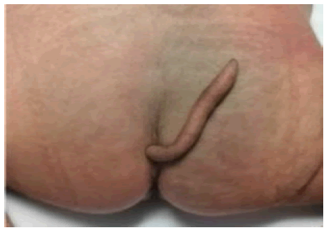
Upon evaluation, a cutaneous appendage that originated from the sacrococcygeal region and rose over the intergluteal fissure in the midline was discovered. The tail-like structure was 9 cm long, cylindrical in shape, and pointed at the end. Its overall diameter ranged from 3 cm to 2 cm. The structure was soft, covered with skin, and demonstrated spontaneous motion.
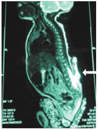
The results of the MRI reveal S1-S2 level spinal bifida (Figure 1). The entire structure was removed during surgery and sent to the pathology laboratory (Figure 2).
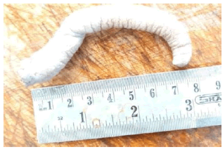
Gross pathologic examination shows a tail-like structure measuring 9 cm in length, had a diameter of 2 cm to 3 cm along its entire length, was cylindrical, and culminated in a tip (Figure 3).
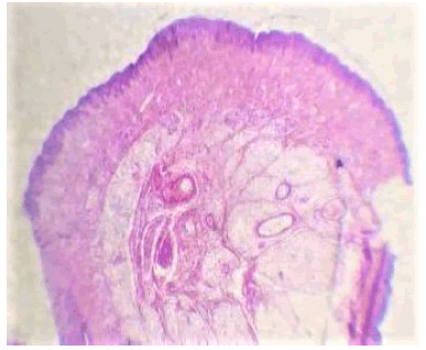
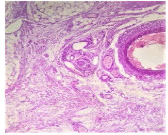
Microscopic examination shows a covering of keratinized stratified squamous epithelium. The dermis contained sweat glands, hair follicles, skeletal muscle, smooth muscles, nerve bundles, adipose tissue, blood vessels, and fibrous mesenchymal tissue (Figure 4 and Figure 5).
DISCUSSION
A tail with 10-12 caudal vertebrae is present in the human embryo during the fifth and sixth weeks of pregnancy. It regresses by having fewer vertebrae and fusing them, leaving only the vestige of a coccyx. A tail typically vanishes by week 8 of pregnancy, however, the precise time varies. A persistent "true" tail is a vestigial feature that most likely develops as a vertebrate remnant of the embryonic tail [3,4].
The most distal end of the typical embryonic tail that is present in the growing human fetus incompletely recedes to form the true human tail. The retention of the embryonic tail in humans may be caused by mutations that increase the WNT3A gene's upregulation [6]
True tails can occur in all races. Although familial occurrences have been described, true human tails are not inherited. In one instance, three generations of females in the same family were born with true human tails. No such family history exists for our newborn patient.
True tails were characterized as looking like a sausage, penis, finger, or stump and were frequently covered in hairy, pigmented skin. Males are twice as likely to develop it as females. Human tails lack the vertebral structures found in the tails of other vertebrates [3-5]. The diameter was between 0.7 cm and 3 cm, and the length varied from 3 cm to 13 cm [4]. In our case, the residual tail is 2 cm to 3 cm in diameter and 9 cm long. It exhibits spontaneous movement.
Histologically, Vestigial tails have central bundles of striated muscle that allow it to move, as well as mature adipose tissue, connective tissue, blood vessels, and nerve fibers. Skin with typical sweat glands and hair follicles covers the surface. Typically, the dermis is thicker than typical [3-6]. Our case shows all vestigial tail histology characteristics. Pseudo-tails are lumbosacral protrusions that resemble true tails on the surface. In addition to being associated with additional lesions such as lipoma, teratoma chondrodystrophy, or parasite fetuses, they present as an abnormal extension of the coccygeal vertebrae and elongation of teratomas, adipose tissue, and cartilage [1,4-6]. It is the most human-like tail form and is linked to the spinal cord and spine abnormalities [1].
Congenital anomalies such as spina bifida, brain and craniofacial diseases, congenital heart disease, anal and vaginal atresia, horseshoe kidney, cleft palate, club foot, and syndactyl can all be accompanied by either a true or a pseudo tail [4-7].
The preferred imaging modality is Magnetic Resonance Imaging (MRI), which has strong specificity for defining anatomy and location as well as excluding other diseases including tethered chord syndrome, occult spina bifida, vertebral arch hypoplasia, myelomeningocele, or meningocele [8-10].
Clinical and radiological examination, followed by simple excision or removal and repair of the underlying lesion if it is accompanied by congenital defects, are essential in the management of human tails [11]. Histopathological examination is necessary to rule out teratomatous components and other related neoplasms.8 To rule out potential problems following surgery, especially in cases involving spinal dysraphism, long-term follow-up is required postoperatively [1,11].
Conclusion
This is an unusual case of a 47-day-old female newborn born to a primiparous mother through spontaneous vaginal birth having a vestige of a human tail and spinal bifida. Imaging is required to rule out any underlying anomalies before surgical therapy, and histopathologic examination is necessary to distinguish between a true tail and a pseudo tail as well as to rule out underlying pathologies.
Declarations
Ethical Approval and Consent to Participate
We sought and obtained written informed consent from the parents of the newborn to publish this article anonymously. This required no further review by the Institutional Review Board (IRB) of Jimma University.
Conflict of Interest
The authors declared no potential conflicts of interest with respect to the research, authorship, and/or publication of this article.
Consent for Publication
Written informed consent was obtained from the parents of the newborn for publication of this case report and any accompanying images. A copy of the written consent is available for review by the Editor-in-Chief of this journal.
Availability of data and materials
Not applicable.
Competing Interests
The authors declare that they have no competing interests.
Funding
This case report received no specific grant or funds.
Author Contribution
All authors have contributed equally.
Acknowledgments
Not applicable.
References
- Donovan, Daniel J., and Robert C. Pedersen. "Human tail with noncontiguous intraspinal lipoma and spinal cord tethering: case report and embryologic discussion." Pediatric neurosurgery, Vol. 41, No. 1, 2005, pp. 35-40.
Google Scholar Crossref - Singh, Deepak Kumar, et al. "The human tail: rare lesion with occult spinal dysraphism-a case report." Journal of Pediatric Surgery, Vol. 43, No. 9, 2008, pp. 41-43.
Google Scholar Crossref - Mukhopadhyay, Biswanath, et al. "Spectrum of human tails: A report of six cases." Journal of Indian Association of Pediatric Surgeons, Vol. 17, No. 1, 2012, p. 23.
Google Scholar Crossref - Dao, Anh H., and Martin G. Netsky. "Human tails and pseudotails." Human pathology, Vol. 15, No. 5, 1984, pp. 449-53.
Google Scholar Crossref - Rueda, Josue, et al. "Human tail in a newborn." Journal of Pediatric Surgery Case Reports, Vol. 76, 2022, p. 102098.
Google Scholar Crossref - Kang, Hyun Koo, et al. "Human tail: a case report." Journal of the Korean Radiological Society, Vol. 47, No. 4, 2002, pp. 419-21.
Google Scholar - Chauhan, S. P. S., et al. "Human tail with spina bifida." British Journal of Neurosurgery, Vol. 23, No. 6, 2009, pp. 634-35.
Google Scholar Crossref - Alashari, Mouied, and Joy Torakawa. "True tail in a newborn." Pediatric dermatology, Vol. 12, No. 3, 1995, pp. 263-66.
Google Scholar Crossref - Daib, Aida, et al. "A rare case of lumbosacrococcygeal mass in newborn: a human tail." Journal of Surgical Case Reports, Vol. 2020, No. 11, 2020, p. 426.
Google Scholar Crossref - Ohhara, Yoshio. "Human tail and other abnormalities of the lumbosacrococcygeal region relating to tethered cord syndrome." Annals of Plastic Surgery, Vol. 4, No. 6, 1980, pp. 507-10.
Google Scholar Crossref - Ahmad, Asrar, Masooma Hasnain Bangash, and Irum Saleem. "Human tail-a rare anatomical mystery: A case report." Journal of Pediatric and Adolescent Surgery, Vol. 1, No. 1, 2020, pp. 44-46.
Google Scholar Crossref

