Research - International Journal of Medical Research & Health Sciences ( 2021) Volume 10, Issue 6
Comparative Analysis of Angiogenic Factor Levels in Platelet-Rich Fibrins Obtained from Titanium and Glass Tubes
Taner Arabaci1 and Mevlut Albayrak2*2Health Services Vocational School, Ataturk University, Erzurum, Turkey
Mevlut Albayrak, Health Services Vocational School, Ataturk University, Erzurum, Turkey, Email: mevlutalbayrak@atauni.edu.tr
Received: 13-May-2021 Accepted Date: Jun 22, 2021 ; Published: 30-Jun-2021
Abstract
In this study, a comparative analysis of angiogenic factor levels from Platelet Rich Fibrin (PRF) obtained from titanium and glass tubes was aimed. Angiotensin-Converting Enzyme (AGE), Interleukin-1a (IL-1a), Platelet-Derived Growth Factor (PDGF), and Interleukin-4 (IL-4) levels of PRF obtained from rats were determined by ELISA (Enzyme-Linked ImmunoSorbent Assay). According to the results obtained; AGE values in the Titanium-Platelet-Rich Fibrin (T-PRF) group were higher than in the control serum group. The mean IL-1a values in the glass tube (G-PRF) group were statistically higher than the other groups. IL-4 levels were found to be high in the G-PRF and T-PRF groups but were not significantly significant. PDGF values were higher in the T-PRF group than in other groups. This study, according to our literature research, is the first study showing the growth factor and cytokine levels in PRFB.
Keywords
Platelet-rich fibrin, Titanium tube, Glass tube, ELISA, Angiogenic factor
Introduction
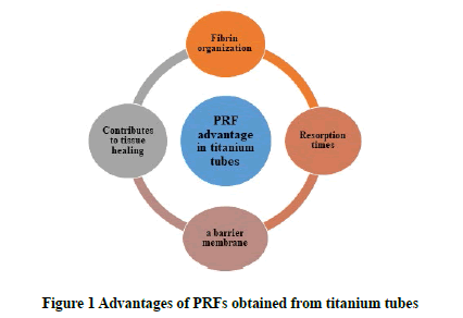
PRF is a second-generation blood-derived autogenous product and has found widespread use in periodontology and other dentistry fields [1]. PRF is also used in facial plastic surgery applications (rhinoplasty, depressive scars, facial volume, acne scars, facial plastic surgery, etc.) [2,3]. It has been shown to contribute to the healing of non-healing lower extremity wounds, and the repair of joint deformations [4,5]. Glass tubes were used to produce this product developed by Choukroun in 2001 [6,7]. However, in the following years, considering the activating properties of titanium on platelets, titanium tubes, which were developed for the first time by Turkish scientists, came into use [8]. It has been shown that the fibrin organization and resorption times of PRFs obtained from titanium tubes have advantageous properties compared to PRFs obtained from glass tubes [8]. PRF functions as a barrier membrane, especially in dental surgery, as well as contributes to tissue healing through platelet-derived angiogenic biomodulators (Figure 1) [9-12].
Platelet-rich plasma is a blood product used to increase tissue healing [13]. It is obtained by centrifugation to concentrate blood platelets drawn from the individual’s peripheral vein. PRP is divided into subgroups depending on fibrin and leukocyte content: These; Leukocyte and Platelet Rich Plasma (L-PRP), Plasma Rich Growth Factor (PRGF), Pure Platelet Rich Plasma (PPRP) [11,13].
Platelets not only prevent blood loss, but also stimulate tissue regeneration, release growth factors and cytokines, triggering angiogenesis and immune response, and increasing collagen synthesis [14]. AGE has an important role in the physiology of blood vessels and the inflammatory process [15]. AGE exerts pro-inflammatory effects by increasing the formation of both Reactive Oxygen Species (ROS) and the synthesis of cytokines called interleukins [16]. During tissue regeneration, PDGF plays a role in both tissue repair and stimulation of cell proliferation as well as extracellular cell proliferation and differentiation [12,17,18].
Platelets are usually immobile and become active as a result of direct contact with collagen exposed to the bloodstream after endothelial damage [19]. Activated platelets, PDGF, and bioactive proteins play essential roles in many stages of hemostasis, including vasoconstriction, leukocyte uptake, and vascular repair [20]. PDGF is produced primarily upon injury and plays a role in all stages of wound healing PDGF in combination with Interleukin-1 (IL-1) draws neutrophils into the wound area to remove contaminating bacteria [9,12]. IL-1 family is the largest family of interleukins and IL-1 cytokines induce strong inflammatory responses [21,22].
Platelet-rich fibrin is produced from the patient’s venous blood in a single centrifugation step without the use of additional anticoagulants and at the same time, PRP consists of autologous plasma with a platelet concentration five times higher than basal level due to an extraction and concentration process [23,24].
They reported that the silica contact of PRF obtained from glass tubes with silica activator is unavoidable and these particles can reach the patient when the product is used for treatment [4].
In a study by the team that developed T-PRF with their titanium tube discoveries, they showed that silica particles were observed in G-PRF while not in T-PRF [25].
They demonstrated that T-PRF gives better results in bone healing and soft tissue compared to PRF obtained with glass tubes and that T-PRF can induce new bone formation with new connective tissue in a rabbit wound healing model within 30 days of treatment [8].
This study aims to quantitatively compare the PRF obtained from newly used titanium tubes and the PRF obtained from glass tubes and Angiogenic Biomodulators (AG, PDGF, IL-1a, IL-4) obtained from glass tubes with the ELISA method.
Materials and Methods
The study was carried out by obtaining the permission certificate number 25330273-929-E.1700151764 from Ataturk University Experimental Animals Ethics Committee. A total of 27 rats weighing 220 g-260 g, aged 12-14 weeks old were used in this study. The animals were obtained from Ataturk University Animal Laboratory (Erzurum, Turkey). AGE, PDGF, IL-1a, and IL-4 ELISA kits were purchased from Biocompare company. Biotech, Epoch Microplate Spectrophotometer device was used for ELISA analysis.
Sample Preparation
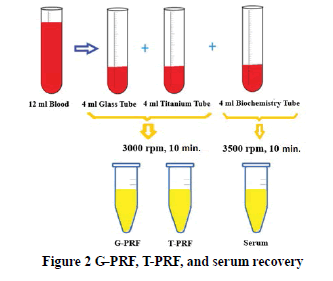
To generate PRF, 12 ml of blood from each rat were divided into three groups of 4 ml. Rats are anesthetized using Thiopental sodium. After the rats were put to sleep, the thorax of each rat was opened and 12 ml of blood was taken directly from their hearts with a 20 ml injector. Then the blood obtained was transferred to a 4 ml glass tube, 4 ml titanium tube, and 4 ml biochemistry tube for serum recovery. The blood in glass and titanium tubes was immediately centrifuged at 3000 rpm and the blood in biochemistry tubes at 3500 rpm for 10 minutes. PRFs obtained in glass and titanium tubes and serums obtained in biochemistry tubes were transferred to microcentrifuge tubes (Figure 2). IL-1a, IL-4, AGE, and PDGF levels were determined from all samples by performing the procedures defined by the manufacturer by the ELISA method.
Statistical Analysis
The data were analyzed using SPSS version 18 statistical software (SPSS Inc., Chicago, IL, USA). Paired t-test and t-test were used to compare continuous variables. Values of p<0.05 were considered statistically significant.
Result and Discussion
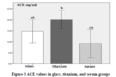
When the AGE results of our study were evaluated; AGE values were found to be significantly higher in the T-PRF group compared to the control serum group (p<0.05), but no difference was observed between the other groups (p>0.05). As a result, the high AGE value in the titanium tube we found makes sense when we consider that AGE has an important role in the inflammatory process and plays a role in platelet activation.
PRF is a biological material that has been used routinely in many areas. However, there has not been a large-scale study on acquisition techniques in the literature. This study is the first study investigating growth factor and cytokine levels in PRFs obtained in glass and titanium tubes.
Biological materials with anti-inflammatory effects and rich in growth factor play an important role in periodontal operations [26]. In periodontal treatment, T-PRF is more successful in periodontal healing than traditional methods [27].
In Figure 3, AGE values were found to be significantly higher in the T-PRF group compared to the control serum group (p<0.05), but there was no difference when compared with the other groups (p>0.05).
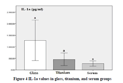
In Figure 4, when IL-1a is evaluated; the mean IL-1a values in the G-PRF group were statistically significantly higher than the other groups (p<0.05). Although the data in the T-PRF group increased compared to the data in the control group, this was not statistically significant (p>0.05).
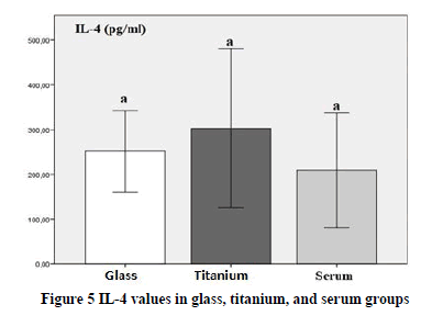
In Figure 5; IL-4 values were found to be high in G-PRF and T-PRF groups, but were not statistically significant (p>0.05). Although IL-4 level was higher in the T-PRF group compared to G-PRF, no statistically significant difference was found (p>0.05).
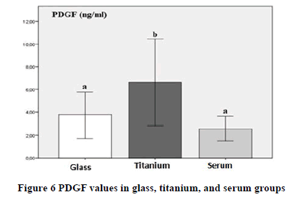
In Figure 6; PDGF values were significantly higher in the T-PRF group compared to other groups (p<0.05). However, PDGF values in G-PRF were found to be similar to the values in control serum (p>0.05).
M. Tunali, et al. showed that titanium tubes are more effective than glass tubes in activating platelets and that T-PRF provides a tighter fibrin network structure than silica in platelet activation [25].
When IL-1a is evaluated; the mean IL-1a values in the G-PRF group were statistically significantly higher than the other groups (p<0.05). Although an increase was observed in the T-PRF group compared to the control group, this was not statistically significant (p>0.05).
Platelet α-granules contain a variety of pro-and anti-angiogenic proteins [28]. Platelet-Derived Growth Factor (PDGF), Hepatocyte Growth Factor (HGF), Epidermal Growth Factor (EGF), Vascular Endothelial Growth Factor (VEGF), Fibroblast Growth Factor (FGF), and Insulin-like Growth Factor (IGF) growth stored in platelet α-granules are among the factors. These angiogenic activators collectively contribute to vascular wall permeability, participation, proliferation, and growth of endothelial cells and fibroblasts [29]. In our study, the values of PDGF, one of these growth factors, were significantly higher in the T-PRF group compared to the other groups (p<0.05). However, PDGF values in G-PRF were found to be similar to the values in control serum (p>0.05). Composed of numerous growth factors and cytokines, PRF is a platelet concentrate that can promote tissue regeneration and repair [30]. J.Kim, et al. showed that the mechanism by which PRF stimulates bone healing may be that platelets are involved in the early diagnosis of bone disease and initiate the repair response by releasing PDGF, Insulin-like Growth Factors (IGF) [31]. In our study, it is a very significant data that PDGF, which has important contributions to periodontal healing, has higher values in T-PRF than the PRF obtained in a glass tube.
The level of IL-4, which is an anti-inflammatory cytokine, was also higher in the T-PRF group, although it was not statistically important (p>0.05). The primary role of IL-4 in inflammation is to support healing, and the presence of this cytokine is sufficient to initiate angiogenesis [32]. Jubert, et al. reported that PRP reduced pain and led to a more effective and lasting functional improvement compared to the placebo substance [33]. The surface of the titanium tubes is often covered by TiOx and/or TiO2 layers [34]. The effect of titanium tubes on the growth factor is the presence of nanobubbles. One of the factors regarding the tubes having different materials is their hydrophilicity/hydrophobicity [35].
Conclusion
According to the results, it has been observed that T-PRF has higher levels of growth factor and cytokine compared to G-PRF and can contribute to periodontal healing. In addition, considering that T-PRF increases bone structure and accelerates healing, it becomes clear that this study is of great importance. When the results of the studies in the literature are evaluated, it is predicted that T-PRF can be used as an advantageous method, especially in surgical applications, in terms of both easy obtaining and low cost. According to our literature research, this study is the first study to show growth factors and cytokine levels in PRFs. We think that our study will guide new studies on T-PRF where other growth factors and cytokines will be compared to create a standard protocol in the future.
Declarations
Conflicts of Interest
The authors declared no potential conflicts of interest concerning the research, authorship, and/or publication of this article.
Acknowledgment
We would like to thanks Ataturk University Scientific Research Projects Coordination Unit for supporting our study with the project number THD-2017-6108.
References
- Eskan, Mehmet Akif, and Henry Greenwell. "Theoretical and clinical considerations for autologous blood preparations: Platelet-rich plasma, fibrin sealants, and plasma-rich growth factors."Clinical Advances in Periodontics,Vol. 1, No. 2, 2011, pp. 142-53.
- Sclafani, Anthony P. "Safety, efficacy, and utility of platelet-rich fibrin matrix in facial plastic surgery."Archives of Facial Plastic Surgery,Vol. 13, No. 4, 2011, pp. 247-51.
- Braccini, F., and D. M. Dohan. "The relevance of Choukroun's Platelet Rich Fibrin (PRF) during facial aesthetic lipostructure (Coleman's technique): Preliminary results."Journal of Laryngology-otology-rhinology,Vol. 128, No. 4, 2007, pp. 255-60.
- O'Connell, Sean M., et al. "Autologous platelet-rich fibrin matrix as cell therapy in the healing of chronic lower-extremity ulcers."Wound Repair and Regeneration,Vol. 16, No. 6, 2008, pp. 749-56.
- Haleem, Amgad M., et al. "The clinical use of human culture-expanded autologous bone marrow mesenchymal stem cells transplanted on platelet-rich fibrin glue in the treatment of articular cartilage defects: A pilot study and preliminary results."Cartilage,Vol. 1, No. 4, 2010, pp. 253-61.
- Choukroun, Joseph, et al. "Platelet-Rich Fibrin (PRF): A second-generation platelet concentrate. Part IV: Clinical effects on tissue healing."Oral Surgery, Oral Medicine, Oral Pathology, Oral Radiology, and Endodontology,Vol. 101, No. 3, 2006, pp. e56-60.
- Borie, Eduardo, et al. "Platelet-rich fibrin application in dentistry: A literature review."International Journal of Clinical and Experimental Medicine,Vol. 8, No. 5, 2015, pp. 7922-29.
- Tunali, Mustafa, et al. "In vivo evaluation of Titanium-prepared Platelet-Rich Fibrin (T-PRF): A new platelet concentrate."British Journal of Oral and Maxillofacial Surgery,Vol. 51, No. 5, 2013, pp. 438-43.
- Hantash, Basil M., et al. "Adult and fetal wound healing."Frontiers in Bioscience,Vol. 13, No. 1, 2008, pp. 51-61.
- Gurbuzer, Bahadir, et al. "Scintigraphic evaluation of osteoblastic activity in extraction sockets treated with platelet-rich fibrin."Journal of Oral and Maxillofacial Surgery,Vol. 68, No. 5, 2010, pp. 980-89.
- Ehrenfest, David M. Dohan, et al. "Classification of platelet concentrates (Platelet-Rich Plasma-PRP, Platelet-Rich Fibrin-PRF) for topical and infiltrative use in orthopedic and sports medicine: current consensus, clinical implications and perspectives."Muscles, Ligaments and Tendons Journal,Vol. 4, No. 1, 2014, pp. 3-9.
- Anitua, Eduardo, Ander Pino, and Gorka Orive. "Opening new horizons in regenerative dermatology using platelet-based autologous therapies."International Journal of Dermatology,Vol. 56, No. 3, 2017, pp. 247-51.
- Choukroun, Joseph, et al."An opportunity in paro-implantology: The PRF."ImplantodonticsVol. 42, No. 55, 2001, p. e62.
- Piccin, Andrea, et al. "Platelet gel: A new therapeutic tool with great potential."Blood Transfusion,Vol. 15, No. 4, 2017, pp. 333-40.
- Jin, Sheng-Yu, et al. "Association of angiotensin converting enzyme gene I/D polymorphism of vitiligo in Korean population."Pigment Cell Research,Vol. 17, No. 1, 2004, pp. 84-86.
- Takahashi, Tomosaburo, et al. "Participation of reactive oxygen intermediates in the angiotensin II-activated signaling pathways in vascular smooth muscle cells."Annals of the New York Academy of Sciences,Vol. 902, No. 1, 2000, pp. 283-87.
- Greenhalgh, David G. "The role of growth factors in wound healing."Journal of Trauma and Acute Care Surgery,Vol. 41, No. 1, 1996, pp. 159-67.
- De Pascale, Maria Rosaria, et al. "Platelet derivatives in regenerative medicine: An update."Transfusion Medicine Reviews,Vol. 29, No. 1, 2015, pp. 52-61.
- Amable, P. R., et al. "Platelet-rich plasma preparation for regenerative medicine: Optimization and quantification of cytokines and growth factors."Stem Cell Research Therapy,Vol. 4, 2013, 67.
- Blair, Price, and Robert Flaumenhaft. "Platelet α-granules: Basic biology and clinical correlates."Blood Reviews,Vol. 23, No. 4, 2009, pp. 177-89.
- Kwak, Areum, et al. "Intracellular interleukin (IL)-1 family cytokine processing enzyme."Archives of Pharmacal Research,Vol. 39, No. 11, 2016, pp. 1556-64.
- Palomo, Jennifer, et al. "The interleukin (IL)-1 cytokine family-Balance between agonists and antagonists in inflammatory diseases."Cytokine,Vol. 76, No. 1, 2015, pp. 25-37.
- Kubesch, Alica, et al. "A low-speed centrifugation concept leads to cell accumulation and vascularization of solid platelet-rich fibrin: An experimental study in vivo."Platelets,Vol. 30, No. 3, 2019, pp. 329-40.
- Moreno, Raquel, Alonso Herreros JM, and A. Villimar. "Methods to obtain platelet-rich plasma and osteoinductive therapeutic use."Hospital Pharmacy: Official Scientific Expression Body of the Spanish Society of Hospital Pharmacy,Vol. 39, No. 3, 2015, pp. 130-36.
- Tunali, Mustafa, et al. "A novel platelet concentrate: Titanium-prepared platelet-rich fibrin."BioMed Research International,Vol. 2014, 2014.
- Dm, Dohan Ehrenfest, L. Rasmusson, and T. Albrektsson. "Classification of platelet concentrates: from Pure Platelet-Rich Plasma (P-PRP) to Leucocyte-and Platelet-Rich Fibrin (L-PRF)."Trends in Biotechnology,Vol. 27, No. 3, 2009, pp. 158-67.
- Arabaci, Taner, and Mevlut Albayrak. "Titanium-prepared platelet-rich fibrin provides advantages on periodontal healing: A randomized split-mouth clinical study."Journal of Periodontology,Vol. 89, No. 3, 2018, pp. 255-64.
- Kisucka, Janka, et al. "Platelets and platelet adhesion support angiogenesis while preventing excessive hemorrhage."Proceedings of the National Academy of Sciences,Vol. 103, No. 4, 2006, pp. 855-60.
- Weltermann, Ansgar, et al. "Large amounts of vascular endothelial growth factor at the site of hemostatic plug formation in vivo."Arteriosclerosis, Thrombosis, and Vascular Biology,Vol. 19, No. 7, 1999, pp. 1757-60.
- Alsousou, J., et al. "The role of platelet-rich plasma in tissue regeneration."Platelets,Vol. 24, No. 3, 2013, pp. 173-82.
- Kim, Jin, Yooseok Ha, and Nak Heon Kang. "Effects of growth factors from platelet-rich fibrin on the bone regeneration."Journal of Craniofacial Surgery,Vol. 28, No. 4, 2017, pp. 860-65.
- Hayashi, Yoshinori, et al. "Interferon-γ and interleukin-4 inhibit interleukin 1β-induced delayed prostaglandin E2 generation through suppression of cyclooxygenase-2 expression in human fibroblasts."Cytokine,Vol. 12, No. 6, 2000, pp. 603-12.
- Joshi Jubert, Nayana, et al. "Platelet-rich plasma injections for advanced knee osteoarthritis: A prospective, randomized, double-blinded clinical trial."Orthopaedic Journal of Sports Medicine,Vol. 5, No. 2, 2017.
- Akhavan, Omid, and Elham Ghaderi. "Flash photo stimulation of human neural stem cells on graphene/TiO 2 heterojunction for differentiation into neurons."Nanoscale,Vol. 5, No. 21, 2013, pp. 10316-26.
- Jannesari, Marziyeh, Omid Akhavan, and Hamid R. Madaah Hosseini. "Graphene oxide in generation of nanobubbles using controllable microvortices of jet flows."Carbon,Vol. 138, 2018, pp. 8-17.

