Research - International Journal of Medical Research & Health Sciences ( 2021) Volume 10, Issue 7
Antioxidant Action of Green Tea Extract in Alloxan-Induced Diabetic Albino Rats: An Experimental Laboratory-Based Study
Ali Hassan A. Ali1,2*, Ali Yaqoub Alali1, Rasheed A. Alhajri1, Abdulrahman M. Aldawsari1, Ammar H. AlHeseni1 and Nasser S. Aldosari12Anatomy Department, Faculty of Medicine, Al-Azhar University, Cairo, Egypt
Ali Hassan A. Ali, Anatomy Department, College of Medicine, Prince Sattam Bin Abdulaziz University, KSA, Email: alihassan3750@yahoo.com
Abstract
Background: Green tea extract can regulate blood sugar and encourage weight loss due to its high antioxidant content. Several studies have shown that green tea extract can improve general health and may help protect against many diseases. Most patients with diabetes are associated with poor fertility. Aim of the work: The present study aimed to evaluate the importance of green tea extract supplementation in reducing metabolic abnormalities associated with alloxan-induced diabetes in male albino rats. Material and Methods: This study was conducted on thirty male albino rats with an average weight of 100 grams-110 grams. The rats were divided equally into three groups that included control, diabetic and diabetic took green tea extracts orally (50 mg/Kg/day) for three weeks. A single dose of alloxan (120 mg/kg body weight) was used to induce diabetes in rats. Diabetic rats were given 50 mg/kg body weight green tea extracts orally for thirty days twice per day. The morphological and histological structures of the testis were compared in different groups of rats. Results: It was revealed that the morphological testicular changes observed in diabetic groups were significantly improved after treatment with green tea compared to the control group. Conclusion: These results prove that green tea improves oxidative damage caused by diabetes in the testis of male rats.
Keywords
Testis, Green tea, Antioxidant, Alloxan, Albino rat
Introduction
Diabetes mellitus is one of the environmental factors that damage the testis and thus affects fertility in men [1]. Worldwide, diabetes is one of the most common metabolic disorders. It is associated with disturbances in the metabolism of carbohydrates, fat, and protein [2]. Experimentally, it was found that the induction of diabetes in male rats is associated with a change in the function of the reproductive system [3,4]. It has been shown that induction of diabetes affects the testicular functions due to insulin deficiency and consequent impairment of insulin regulatory action on the Sertoli and Leydig cells [5]. In diabetic cases there are several studies showed changes in the structure of the reproductive system [6-8].
Diabetes mellitus is a multifactorial disease with a deficiency in Reactive Oxygen Species (ROS) scavenging enzymes [9]. The prime cause of a number of long-term complications of diabetes is chronic hyperglycemia. The most important source of free radicals is protein glycation caused by hyperglycemia. ROS can directly cause molecular as well as cellular damage by activating many cellular stress-sensitive pathways, which lead to diabetes complications [10]. Several secondary metabolites of the plant have shown antioxidant potential and improved effect on damage-induced oxidative stress in diabetes [11].
Traditional medicine practices are responsible for a neutral role in primary health care despite the accessibility of modern medicine [12].
Dietary antioxidants, in general, are often safe substances found in medicinal plants and have attractive effects in complementary medicine including an important effect in reducing oxidative stress [13,14]. The polyphenols and catechins that make up large groups of nutritional antioxidants are especially found in green tea [15,16]. In addition to their antioxidant effect, these nutrients have anti-diabetic, antihypertensive, antioxidative, anti-inflammatory, and antifungal functions [17,18]. Therefore, green tea consumption is associated with a lower mortality rate especially from cardiovascular strokes [19]. The mechanism of protection exerted by green tea consumption against chronic diseases is unclear, however, it had been suggested that the protective role of green tea consumption with these diseases may be invoked to the antioxidant action of its polyphenols and catechins [16,20,21].
The present study aimed to investigate the anti-diabetic effects of green tea aqueous extract on testis in male albino rats with diabetes mellitus induced by alloxan.
Materials and Methods
Green tea extract was obtained as 300 mg tablets synthesized by Tecno Med Company-KSA. The tablets were crushed and the required amount was dissolved in distilled water. It left cool to room temperature then filtered. The extract was daily prepared and stored in glass containers in a refrigerator.
Animals
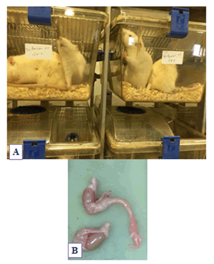
In this experimental study, 30 adults, healthy, male Wistar albino rats (300 grams-350 grams, 3 months old) were used. They were gained from an animal house of the College of Pharmacy, Prince Sattam Bin Abdulaziz University, KSA. They were kept under controlled standard animal housing situations and moisture with access to food and water ad libitum at the Animal Care Facility at the Department of Clinical Pharmacy at Prince Sattam bin Abdulaziz University (Figure 1). The pellet diet is composed of 23% protein, 5% lipids, 4% crude fiber, and 55% nitrogen-free extract. The rats were kept under control for approximately 2 weeks before the start of the experimentation to allow acclimatization. Diabetes mellitus was induced in animals except for the control group with a single dose of alloxan (120 mg/kg B.W. dissolved in saline), injected intraperitoneally to induce diabetes mellitus [22].
Ethical Considerations
All procedures that were performed in this study that involved animal models were in accordance with the ethical standards of the Institutional Ethics Committee of Prince Sattam bin Abdulaziz University and the Animal House Committee (IRB, PSAU-2020 ANT 1/39PI).
Experimental Design
Three experimental groups, ten rats for each, were used as follows:
Group I (Control group): Non-diabetic control rats.
Group II (Diabetic group): Rats were injected intraperitoneally with a single dose of alloxan (120 mg/kg dissolved in saline solution).
Group III (Diabetic group+green tea): Diabetic rats were injected also intraperitoneally with a single dose of alloxan (120 mg/kg dissolved in saline solution). Then, treated orally with green tea extract 50 mg/kg body wt. twice/ day green tea extracts orally for 30 days.
After sacrificing animals, their testes were processed, stained, and examined microscopically.
Histological and Histochemical studies: After one month, the rats of all groups were sacrificed and small pieces of the testis were taken for the histological and histochemical studies. The specimens were prepared by fixation in 10% neutral buffered formalin solution and Carnoy’s fluid. For the histological study, paraffin sections were stained with Harris’s Hematoxylin and Eosin (H and E) [23]. Mallory’s trichrome stain was used in paraffin sections for the detection of collagen fibers. Paraffin sections of 5 μm thickness were prepared and stained with Feulgen stain for histochemical study [23]. Then, all the stained sections were examined by light microscope, photographed and all the differences detected between the three groups were discussed at the level of the microscopic findings.
For the immunohistochemical study, the sections of testes were deparaffinized with xylene, then antigen retrieval by heating in citrate buffer (10 mM, 20 min). This was followed by endogenous peroxidase blocking in 3% H2O2 for 10 min. Then and incubated with anti-caspase-3 (1:100; Abcam, Ab4051). The sections were incubated with the related secondary antibodies after washing the slides with phosphate-buffered saline, at room temperature for one hour. Next, 3-amino-9-ethyl carbazole, a chromogen was detected.
Statistical Analyses
Statistical analyses were performed using the ANOVA test. A p-value<0.05 was considered statistically significant.
Results
Histological Results
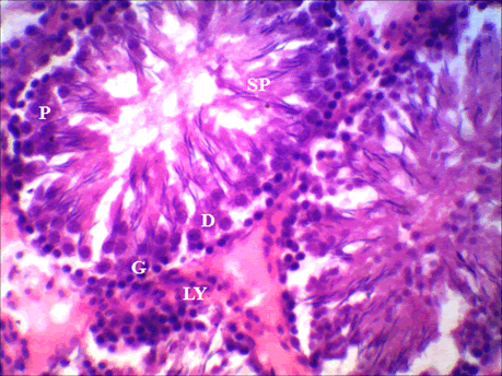
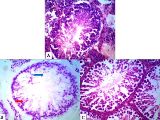
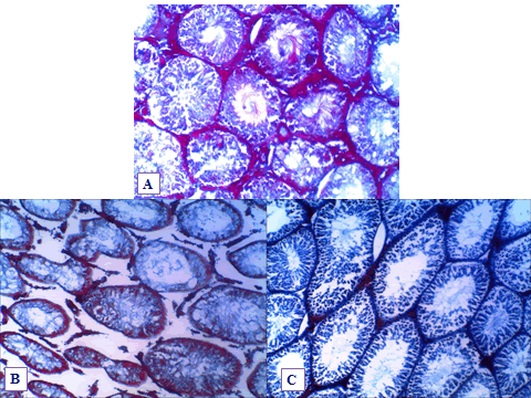
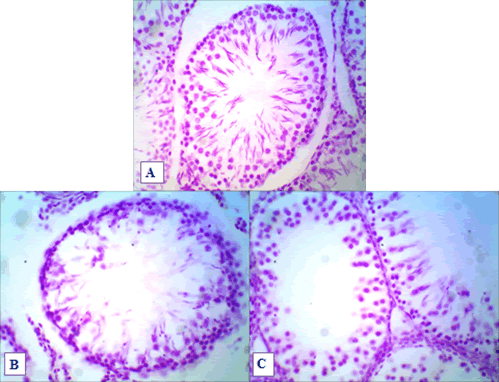
The control adult albino rat testis showed several seminiferous tubules cut in various planes of sections. The seminiferous tubules were highly convoluted and lined germ cells in various stages of spermatogenesis and spermiogenesis which are collectively referred to as spermatogenic series. Non-germ cells called Sertoli cells were well preserved. In the interstitial spaces between the tubules, endocrine cells called Leydig cells were found singly or in groups in the supporting tissue (Figure 2). The histological examination of testicular tissue in the untreated diabetic rats showed irregularity of the seminiferous tubules shapes with a significant reduction in its diameters compared to the control group. In addition, the germinal epithelium showed a clear disruption of the germinal epithelium with abnormal cellular attachment. Furthermore, the spermatogonia cells were the main type of cells seen (Figure 3, and Table 1). Multinucleated cells with two or three nuclei were also detected in seminiferous tubules. Moreover, the interstitial connective tissue had an amorphous material with marked destruction of the connective tissues with subsequent widening of the interstitial spaces (Figure 4). The green tea-treated group showed a significant recovery of the diameters of the seminiferous tubules compared to the diabetic group (Figure 3). It also showed a significant recovery of the interstitial spaces to normal levels with a considerable increase in the amount of the collagen fibers compared to the diabetic group (Figure 4).
| Parameters Study Groups | Mean diameters of the seminiferous tubules | Mean thickness of the seminiferous tubular interstitial diameters | Mean area percentage of the collagen fibers of the seminiferous tubular interstitial diameters | Mean number of the caspase-3+ve cells of the seminiferous tubular germinal cells |
|---|---|---|---|---|
| Group I (Control) | 177.12 ± 14.92 | 94.86 ± 6.57 | 152.03 ± 23.25 | 12.00 ± 1.84 |
| Group II (Diabetic) | 126.63 ± 32.18 | 180.70 ± 21.4 | 65.87 ± 15.85 | 34.71 ± 2.87 |
| Group III (Green tea) | 159.13 ± 19.99 | 66.65 ± 11.07 | 67.93.11 ± 10.72 | 32.82 ± 2.65 |
Figure 4. Histological light photomicrograph of testis sections (Mallory’s trichrome 200×) from the control group shows a normal distribution of collagen fibers in the Interstitial Tissue (IT) around the seminiferous tubules; B): A section form untreated diabetic rat shows marked reduction of collagen fibers in the IT around the seminiferous tubules and increase the diameter of the interstitial spaces; C): Light photomicrograph of a section in a rat testicular tissue treated with green tea shows the mild distribution of collagen fibers in the I.T around the seminiferous tubules in comparison to the control group
Immunohistochemical Study
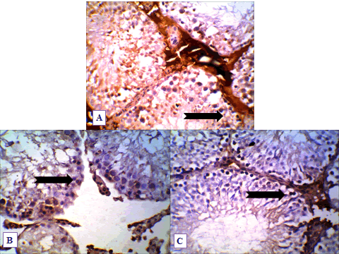
The immunohistochemical examination of the testicular tissue for the control group demonstrated mild expression of Caspase-3 immunostaining with few caspase-3+ve cells in the seminiferous tubules (Figure 5, and Table 1). On the other hand, in the diabetic group, a marked significant increase in the number of caspase-3+ve cells was observed in the seminiferous tubules in comparison to the control ones (Figure 6, and Table 1). The green tea-treated group showed a marked significant increase in the number of caspase-3+ve cells was observed in the seminiferous tubules in comparison to the control ones (Figure 6, and Table 1).
Figure 5. Histological light photomicrograph of testis sections (Feulgen stain 400×) from the control group. The Seminiferous Tubules (STs) have shown a normal association of germ cells; B): A section in a rat testicular tissue from untreated diabetic rat shows marked reduction, pyknosis, disorganization, and depletion of the germinal epithelium in the STs; C): A section in a rat testicular tissue treated with green tea extract shows the marked recovery of the germinal epithelium to the normal architecture
Figure 6. Histological light photomicrograph of testis sections (Caspase-3 immunostaining 400×) from the control group. The S.Ts germinal cells show mild expression of Caspase-3 immunostaining (black arrow); B): The seminiferous tubules germinal cells of untreated diabetic rat show marked expression of Caspase-3 immunostaining (black arrow); C): Light photomicrograph of a section in a rat testicular tissue treated with green tea shows marked expression of Caspase-3 immunostaining (black arrow) in the S.Ts germinal cells
Discussion
The results of the current study showed that green tea has significant protective effects against apoptotic changes. This protective effect may be due to their antioxidant properties.
Free radicals may be caused by tissue injury associated with diabetes and its subsequent complications [24].
Tea is one of the most highly consumed beverages worldwide, especially green tea, which contains phenolic compounds including epigallocatechin gallate, catechins, epicatechin gallate. The antioxidant activity of green tea polyphenols and, more recently, the pro-oxidant effects of these compounds, leading to indirect antioxidant effects, have also been suggested as potential mechanisms for the prevention of cancer [25,26].
Another study showed, green tea extract significantly increased levels of serum insulin in diabetic rats, and green tea extract (combined with ginseng roots) protected the cells in the islets of Langerhans [27].
Other studies have indicated increased thickness of seminiferous tubules and germ cell depletion in both diabetic humans and rats [28,29].
Various herbal extracts or derivatives with high antioxidant activity are useful for diabetes treatment and other metabolic syndromes [30]. Accordingly, antioxidant therapy is one of the major strategies for treating diabetes [30].
Antioxidant defense by antioxidant enzymes is a protective mechanism against oxidative stress that eliminates harmful reactive oxygen species in male reproductive organs. It also plays an important role in preserving reproductive functions [31].
Green tea extract helps to improve diabetic renal changes mainly by correcting oxidative stress as shown in another previous study [32].
Many studies support the benefits of antioxidants in protecting the testis from oxidative stress [33-35].
Finally, considerable improvements in the testicular tissue morphological changes that were observed in diabetic groups had been detected after green tea treatment in comparison to the control group.
Conclusion
The current study showed that green tea has antioxidant activities and reduces the side effects of diabetes mellitus on testicular structures.
Declarations
Authors’ Contributions
This work was performed as a collaboration among all of the authors. Ali Hassan A. Ali and Ali Yaqoub Alali participated in the study design and wrote the first draft of the manuscript. Rasheed A. Alhajri, Abdulrahman M. Aldawsari, and Ammar H. AlHeseni collected and processed the samples. Nasser S. Aldosari participated in the study design and performed the statistical analyses. All of the authors read and approved the final manuscript.
Acknowledgments
This publication was supported by the Deanship of Scientific Research at Prince Sattam bin Abdulaziz University, Alkharj, Saudi Arabia.
Availability of Data and Materials
The data are available upon request from the authors.
Ethics Approval
The study was approved by the Ethics Committee of Prince Sattam bin Abdulaziz University Institutional Review Board.
Conflicts of Interest
The authors declared no potential conflicts of interest concerning the research, authorship, and/or publication of this article.
References
- Sebastian, R., and S. C. Raghavan. "Endosulfan induces male infertility." Cell Death & Disease, Vol. 6, No. 12, 2015, p. e2022.
- Moore, Helen, et al. "Dietary advice for treatment of type 2 diabetes mellitus in adults." Cochrane Database of Systematic Reviews, Vol. 2, 2004.
- Kuhn-Velten, N., D. Waldenburger, and W. Staib. "Evaluation of steroid biosynthetic lesions in isolated leydig cells from the testes of streptozotocin-diabetic rats." Diabetologia, Vol. 23, No. 6, 1982, pp. 529-33.
- Morimoto, S., et al. "Protective effect of testosterone on early apoptotic damage induced by streptozotocin in rat pancreas." Journal of Endocrinology, Vol. 187, No. 2, 2005, pp. 217-24.
- Ballester, Joan, et al. "Insulin‐dependent diabetes affects testicular function by FSH‐and LH‐linked mechanisms." Journal of Andrology, Vol. 25, No. 5, 2004, pp. 706-19.
- Cai, Lu, et al. "Apoptotic germ-cell death and testicular damage in experimental diabetes: Prevention by endothelin antagonism." Urological Research, Vol. 28, No. 5, 2000, pp. 342-47.
- Sanguinetti, Roberto E., et al. "Ultrastructural changes in mouse Leydig cells after streptozotocin administration." Experimental Animals, Vol. 44, No. 1, 1995, pp. 71-73.
- Oztürk, F., et al. "Histological alterations of rat testes in experimental diabetes." T Kin J Med Sci, Vol. 22, 2002, pp. 173-78.
- Kesavulu, M. M., et al. "Lipid peroxidation and antioxidant enzyme levels in type 2 diabetics with microvascular complications." Diabetes & Metabolism, Vol. 26, No. 5, 2000, pp. 387-92.
- Evans, Joseph L., et al. "Are oxidative stress-activated signaling pathways mediators of insulin resistance and β-cell dysfunction?" Diabetes, Vol. 52, No. 1, 2003, pp. 1-8.
- Ugochukwu, N. H., et al. "The effect of Gongronema latifolium extracts on serum lipid profile and oxidative stress in hepatocytes of diabetic rats." Journal of Biosciences, Vol. 28, No. 1, 2003, pp. 1-5.
- Wazaify, Mayyada, et al. "Complementary and alternative medicine use among Jordanian patients with diabetes." Complementary Therapies in Clinical Practice, Vol. 17, No. 2, 2011, pp. 71-75.
- Hamden, Khaled, et al. "Improvement effect of green tea on hepatic dysfunction, lipid peroxidation and antioxidant defence depletion induced by cadmium." African Journal of Biotechnology, Vol. 8, No. 17, 2009, pp. 4233-38.
- Elzoghby, Rabab R., et al. "Protective role of vitamin C and green tea extract on malathion-induced hepatotoxicity and nephrotoxicity in rats." American Journal of Pharmacology and Toxicology, Vol. 9, No. 3, 2014, p. 177.
- Adel, A. G., et al. "Protective role of green tea on carbon tetrachloride-induced acute hepatotoxicity." Scientific Journal of Faculty of Science (Minufiya University), Vol. 23, 2009, pp. 55-67.
- Higdon, Jane V., and Balz Frei. "Tea catechins and polyphenols: Health effects, metabolism, and antioxidant functions." Critical Reviews in Food Science and Nutrition, Vol. 43, No. 1, 2003, pp. 89-143.
- Heikal, Tarek M., et al. "The ameliorating effects of green tea extract against cyromazine and chlorpyrifos induced liver toxicity in male rats." Changes, Vol. 5, No. 9, 2013, pp. 48-55.
- Al-Attar, Atef M., and Isam M. Abu Zeid. "Effect of tea (Camellia sinensis) and olive (Olea europaea L.) leaves extracts on male mice exposed to diazinon." BioMed Research International, Vol. 2013, 2013.
- Kuriyama, Shinichi, et al. "Green tea consumption and mortality due to cardiovascular disease, cancer, and all causes in Japan: the Ohsaki study." JAMA, Vol. 296, No. 10, 2006, pp. 1255-65.
- Frei, Balz, and Jane V. Higdon. "Antioxidant activity of tea polyphenols in vivo: Evidence from animal studies." The Journal of Nutrition, Vol. 133, No. 10, 2003, pp. 3275S-84S.
- Lotito, Silvina B., and Balz Frei. "Consumption of flavonoid-rich foods and increased plasma antioxidant capacity in humans: Cause, consequence, or epiphenomenon?" Free Radical Biology and Medicine, Vol. 41, No. 12, 2006, pp. 1727-46.
- Malaisse, Willy J. "Alloxan toxicity to the pancreatic B-cell: A new hypothesis." Biochemical Pharmacology, Vol. 31, No. 22, 1982, pp. 3527-34.
- Bancroft, John D., and Marilyn Gamble. "Theory and practice of histology techniques." Churchill Livingstone Elsevier, 2008.
- Lapolla, Annunziata, Domenico Fedele, and Pietro Traldi. "Glyco-oxidation in diabetes and related diseases." Clinica Chimica Acta, Vol. 357, No. 2, 2005, pp. 236-50.
- Hou, Zhe, et al. "Mechanism of action of (−)-epigallocatechin-3-gallate: Auto-oxidation-dependent inactivation of epidermal growth factor receptor and direct effects on growth inhibition in human esophageal cancer KYSE 150 cells." Cancer Research, Vol. 65, No. 17, 2005, pp. 8049-56.
- Butt, Masood Sadiq, and Muhammad Tauseef Sultan. "Green tea: Nature's defense against malignancies." Critical Reviews in Food Science and Nutrition, Vol. 49, No. 5, 2009, pp. 463-73.
- Karaca, Turan, et al. "Effects of extract of green tea and ginseng on pancreatic beta cells and levels of serum glucose, insulin, cholesterol and triglycerides in rats with experimentally streptozotocin-induced diabetes: A histochemical and immunohistochemical study." Journal of Animal and Veterinary Advances, Vol. 9, No. 1, 2010, pp. 102-07.
- Cameron, Don F., Frederick T. Murray, and David D. Drylie. "Interstitial compartment pathology and spermatogenic disruption in testes from impotent diabetic men." The Anatomical Record, Vol. 213, No. 1, 1985, pp. 53-62.
- Sadik, Nermin AH, Mohamed M. El-Seweidy, and Olfat G. Shaker. "The antiapoptotic effects of sulphurous mineral water and sodium hydrosulphide on diabetic rat testes." Cellular Physiology and Biochemistry, Vol. 28, No. 5, 2011, pp. 887-98.
- Samad, Abdus, et al. "Status of herbal medicines in the treatment of diabetes: A review." Current Diabetes Reviews, Vol. 5, No. 2, 2009, pp. 102-11.
- Fujii, Junichi, et al. "Cooperative function of antioxidant and redox systems against oxidative stress in male reproductive tissues." Asian Journal of Andrology, Vol. 5, No. 3, 2003, pp. 231-42.
- Pedraza-Chaverrí, José, et al. "S-allylmercaptocysteine scavenges hydroxyl radical and singlet oxygen in vitro and attenuates gentamicin-induced oxidative and nitrosative stress and renal damage in vivo." BMC Clinical Pharmacology, Vol. 4, No. 1, 2004, pp. 1-13.
- El-Sokkary, Gamal H., et al. "Inhibitory effect of melatonin on products of lipid peroxidation resulting from chronic ethanol administration." Alcohol and Alcoholism, Vol. 34, No. 6, 1999, pp. 842-50.
- Lombardo, Francesco, et al. "The role of antioxidant therapy in the treatment of male infertility: An overview." Asian Journal of Andrology, Vol. 13, No. 5, 2011, pp. 690-97.
- Maneesh, M., et al. "Experimental therapeutic intervention with ascorbic acid in ethanol induced testicular injuries in rats." Indian Journal of Experimental Biology, Vol. 43, 2005, pp. 172-76.

