Case Report - International Journal of Medical Research & Health Sciences ( 2023) Volume 12, Issue 2
A Solitary Trichilemmoma in Uncommon Location Of Vulva: A Rare Case Report
Triveni GS*, Kiran Aggarwal, Prabha Lal, Anuradha Singh, Aprajita Gupta and SanghamitaTriveni GS, Department of Obstetrics and Gynecology Lady Hardinge Medical College, New Delhi, India, Email: drtriveni.gs@gmail.com
Received: 16-Dec-2022, Manuscript No. ijmrhs-23-83889; Editor assigned: 19-Dec-2022, Pre QC No. ijmrhs-23-83889 (PQ); Reviewed: 22-Dec-2022, QC No. ijmrhs-23-83889 (Q); Revised: 07-Feb-2023, Manuscript No. ijmrhs-23-83889 (R); Published: 28-Feb-2023
Abstract
Trichilemmoma is a benign neoplasm with differentiation towards pilosebaceous follicular epithelium or outer root sheath. It often presents with solitary lesions but when multiple can be associated with Cowden syndrome with various risks of neoplasms. It is commonly found in the region of the head and neck. It is often asymptomatic and long-standing. The treatment is optional but malignancy has to be excluded in the rapidly growing lesion. We are hereby presenting the occurrence of trichilemmoma in a rare location over the genital area. Initial biopsy was suggestive of features of trichilemmoma. She underwent total excision due to fear of malignancy and cosmetic reasons. The histopathology findings corroborated with trichilemmoma. The patient is asymptomatic without any local complaints and recurrence on follow-up for 6 months. Trichilemmoma should be kept as a differential diagnosis in the verrucous lesions over the genital skin.
Keywords
Solitary, Trichilemmoma, Vulva, Uncommon
Introduction
Cutaneous adnexal tumors are a group of benign and malignant neoplasms with morphological differentiation towards one of the primary adnexal structures present in the normal skin like hair follicles, sebaceous glands, apocrine glands, and eccrine glands. Trichilemmomas are benign tumors arising from the outer cells of the hair follicle. They are sporadic. Trichilemmoma may occur as solitary or multiple tumors. Solitary trichilemmoma typically presents as a small, single-skin colored papule or nodule on the face. In contrast, multiple trichilemmomas typically exhibit a perinasal and perioral distribution and sometimes can be found in the chest and acral sites. Multiple trichilemmomas serve as a cutaneous marker for Cowden’s disease, a rare genetic autosomal dominant inherited cancer syndrome [1]. Solitary trichilemmoma is a relatively uncommon occurrence in the pelvic region. we are presenting a case of trichilemmoma in the unusual site of female external genital skin over the mons pubis. As per the literature, not many trichilemmomas have been reported so far in this rare location.
Case Report
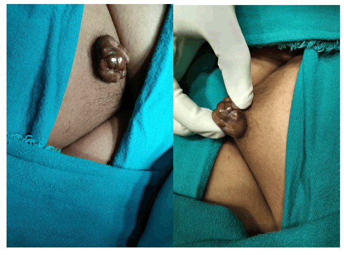
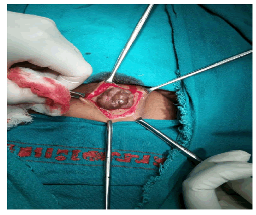
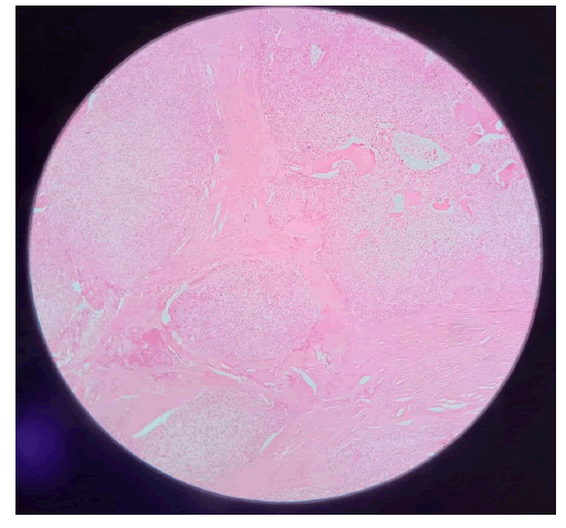
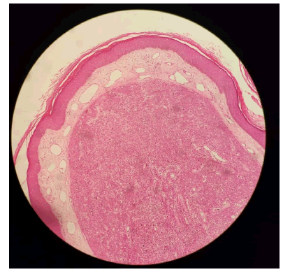
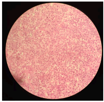
A 42-year-old lady presented to Gynaecology OPD with complaints of swelling over mons pubis for 7 years. This growth was initially pea-sized which had increased to its present size in the last 2 years (Figure 1). The swelling was associated with dryness. It was not associated with pain, redness, itching, or burning sensation. On examination, a variegated cauliflower-like growth of size 3 cm × 4 cm was found on the mons pubis. It was firm in consistency and non-tender with no secondary changes on the skin. She did not have any other features of Cowden syndrome. An initial skin biopsy was done and histopathological findings were suggestive of Trichilemmoma (Figure 2). The patient opted for an excision of mass for cosmetic reasons and fear of malignancy. The decision for wide local excision was made in consultation with the dermatologist and the mass was removed. A biopsy specimen revealed a characteristic folliculocentric tumor with a bulbous profile and stromal clefting seen in the dermis with connection to the epidermis along with basaloid cells and clear cells with hyperkeratosis suggestive of Trichilemmoma. The patient is under follow-up for 6 months without any local complaints or recurrence (Figure 3-5).
Discussion
Cutaneous appendageal tumors often arise from apocrine, eccrine, hair follicles, and sebaceous glands. Trichilemmoma is an epithelial cutaneous tumor resulting from the hamartomatous proliferation of cells from the hair follicle. The multiplication of glycogen-rich clear cells results in the formation of papular or nodular lesions over the skin. It is often seen in the age group of 8 years-80 years [2].
It is often a benign entity but carries malignant potential in long-standing lesions. The lesions can be single or multiple distributed typically over the central face like eyelids, nose, and upper lip but can uncommonly affect other areas like genital skin. Multiple trichilemmomas are a cutaneous marker for an underlying Autosomal Dominant disease called Cowden Syndrome. It is charecterised by multiple trichilemmomas, mucosal papillomas and hamartomas. These individuals have an increased risk of skin, thyroid, breast, endometrium, gastrointestinal and brain malignancies [3,4]. Individuals with multiple Trichilemmomas should be screened for internal malignancies [5]. Though rare, they can also transform into trichilemmal carcinoma.
The microscopic examination displays a lobulated proliferation composed of central pale, periodic acid Schiff positive, diastase sensitive, glycogen-containing cells and peripheral basophilic palisading cells enclosed within an eosinophilic periodic acid Schiff positive basement membrane [6]. The differential diagnosis of trichilemmoma includes verruca vulgaris and hair follicles and epidermal tumors [7]. The trichilemmoma has a desmoplastic variant that can develop on the genital area and may also occur as a secondary neoplasm in the Nevus Sebaceous. Trichilemmoma often requires no treatment. The treatment is usually optional. The biopsy of the lesion confirms the diagnosis and rules out malignancy. The lesion is often removed for aesthetic purposes or when it causes discomfort due to its location over a sensitive area. The approach includes surgical excision or an ablative procedure like cryotherapy, curettage, and carbon dioxide laser therapy [8]. Although a benign entity, the final diagnosis requires a correlation between the clinical and histopathological findings.
A similar case of solitary trichilemmoma on the vulvar region has been reported 72-year-old lady by Lamiaa Hamie et al in 2021. She presented with a 4 mm keratotic verrucous papule of two weeks duration over the right vulvar region. An excision biopsy of the lesion was suggestive of trichilemmoma [9].
Our patient had growth over a genital area and hence had hesitancy in seeking opinion and care for years. Trichilemmoma at such a rare location tends to be missed and go unnoticed by patients themselves due to covering with pubic hairs. So, while doing an internal gynecological examination for any indication, it is prudent to do a thorough local examination of the pelvic region to identify such lesions promptly and act accordingly in time for the patient’s benefit. Trichilemmoma must be included in the differential diagnosis of epithelial lesions arising in this rare location. It is particularly important to rule out invasive malignancies like trichilemmal carcinoma, desmoplastic squamous cell carcinoma, sclerosing basal cell Carcinoma, Sebaceous Carcinoma, clear cell Porocarcinoma, clear cell Hidradenocarcinoma, balloon cell melanoma [10].
Conclusion
The Trichilemmoma can present in the unusual location of the mons pubis and Vulva. It should be considered in the differential diagnosis of verrucous growth in the genital area. The removal can be considered when associated with recent rapid growth, to rule out aggressive cutaneous neoplasms.
Declarations
Conflict of Interest
The authors declared no potential conflicts of interest with respect to the research, authorship, and/or publication of this article.
References
- Brownstein, Martin H., and Lewis Shapiro. "Trichilemmoma: analysis of 40 new cases." Archives of Dermatology, Vol. 107, No. 6, 1973, pp. 866-69.
Google Scholar Crossref - Afshar, Maryam, Robert A. Lee, and Shang I. Brian-Jiang. "Desmoplastic trichilemmoma-a report of successful treatment with Mohs micrographic surgery and a review and update of the literature." Dermatologic surgery, Vol. 38, No. 11, 2012, pp. 1867-71.
Google Scholar Crossref - Brownstein, Martin H., et al. "The dermatopathology of Cowden's syndrome." British Journal of Dermatology Vol. 100, No. 6, 1979, pp. 667-73.
Google Scholar Crossref - Kennedy, Robert A., Selvam Thavaraj, and Salvador Diaz-Cano. "An overview of autosomal dominant tumour syndromes with prominent features in the oral and maxillofacial region." Head and neck pathology, Vol. 11, 2017, pp. 364-76.
Google Scholar Crossref - Gorlin, Robert J., and Heddie O. Sedano. "Stomatologic Aspects of Cutaneous Diseases: Multiple Hamartoma Syndrome (Cowden's Syndrome)." Dermatologic Surgery, Vol. 5, No. 1, 1979, pp. 12-13.
Google Scholar Crossref - Maher, Eamonn E., and Claudia I. Vidal. "Trichilemmoma." Cutis, Vol. 96, No. 2, 2015, pp. 81-106.
Google Scholar - Tellechea, Oscar, et al. "Benign follicular tumors." Anais brasileiros de dermatologia, Vol. 90, 2015, pp. 780-98.
Google Scholar Crossref - Jha, A. K., S. Prasad, and R. Sinha. "Linear trichilemmoma following a blaschkoid pattern: a clinical dilemma." Journal of the European Academy of Dermatology and Venereology, Vol. 30, No. 2, 2016, pp. 299-301.
Google Scholar Crossref - Hamie, Lamiaa, et al. "An enlarging keratotic verrucous papule on the vulva." The gulf journal of Dermatology and Venereology, Vl. 28, No. 1, 2021, pp. 52-54.
Google Scholar - Fronek, Lisa, et al. "A Rare Case of Trichilemmal Carcinoma: Histology and Management." The Journal of Clinical and Aesthetic Dermatology, Vol. 14, No. 6, 2021, p. 25.
Google Scholar

